
|
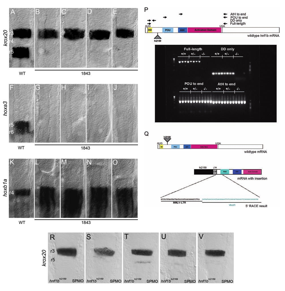
Fig. 3 (A–O) Wild-type (A, F, K) and hnf1b1843 (B–E, G–J, L–O) embryos were assayed for expression of krox20 (A–E), hoxa3 (F–J), and hoxb1a (K–O) by in situ hybridization. All panels are dorsal views of flat-mounted hindbrains with anterior to the top. Rhombomere numbering is indicated in panels (A), (F), and (K). (P) Wild-type (+/+), heterozygous (+/-), and homozygous hnf1bhi2169 mutant (-/-) embryos were assayed for presence of hnf1b transcripts by RT-PCR. Virus integration site and locations of primers used for RT-PCR analysis are indicated in diagram at top. RT-PCR products were resolved by gel electrophoresis, and the resulting gel is shown at the bottom. (Q) Primer extension analysis of virus-hnf1b transcript. Top panel shows structure of wild-type hnf1b mRNA and indicates retroviral insertion site (triangle) in the hnf1bhi2169 allele. Bottom panel shows putative viral-mRNA fusion transcript and indicates sequence obtained from primer extension analysis. (R–V) hnf1bhi2169 embryos injected with an hnf1b spliceblocking morpholino (SPMO) show near-complete loss of krox20 expression in r5 compared to Figure 1B–E.