
Fig. 1
|
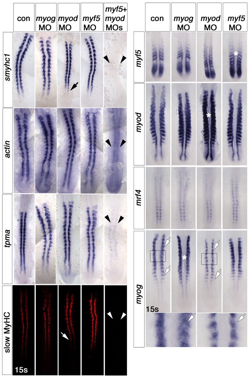
Fig. 1 Myf5 or Myod are required for slow fibre formation. Dorsal flatmounts of zebrafish embryos injected with the indicated MOs and uninjected controls (con), analysed at the 13- to 15-somite stage by whole-mount immunohistochemistry for MyHC (red) or in situ mRNA hybridisation for the indicated mRNAs (blue). Anterior to top. (Left panels) myf5+myod MOs ablated all slow myosin heavy chain (smyhc1 and slow MyHC, arrowheads), but did not completely prevent actin or tpma expression (arrowheads). myod MO slightly delayed smyhc mRNA and MyHC accumulation (arrows), whereas other single MOs had little effect. (Right panels) Single MRF MOs had no effect on adaxial expression of the other MRFs. However, the myod MO reduced myog signal in fast muscle precursors (white arrows). The box region is shown magnified beneath. Note the increase in signal for each MRF caused by its cognate MO (asterisks). s, somites.