
Fig. 4
|
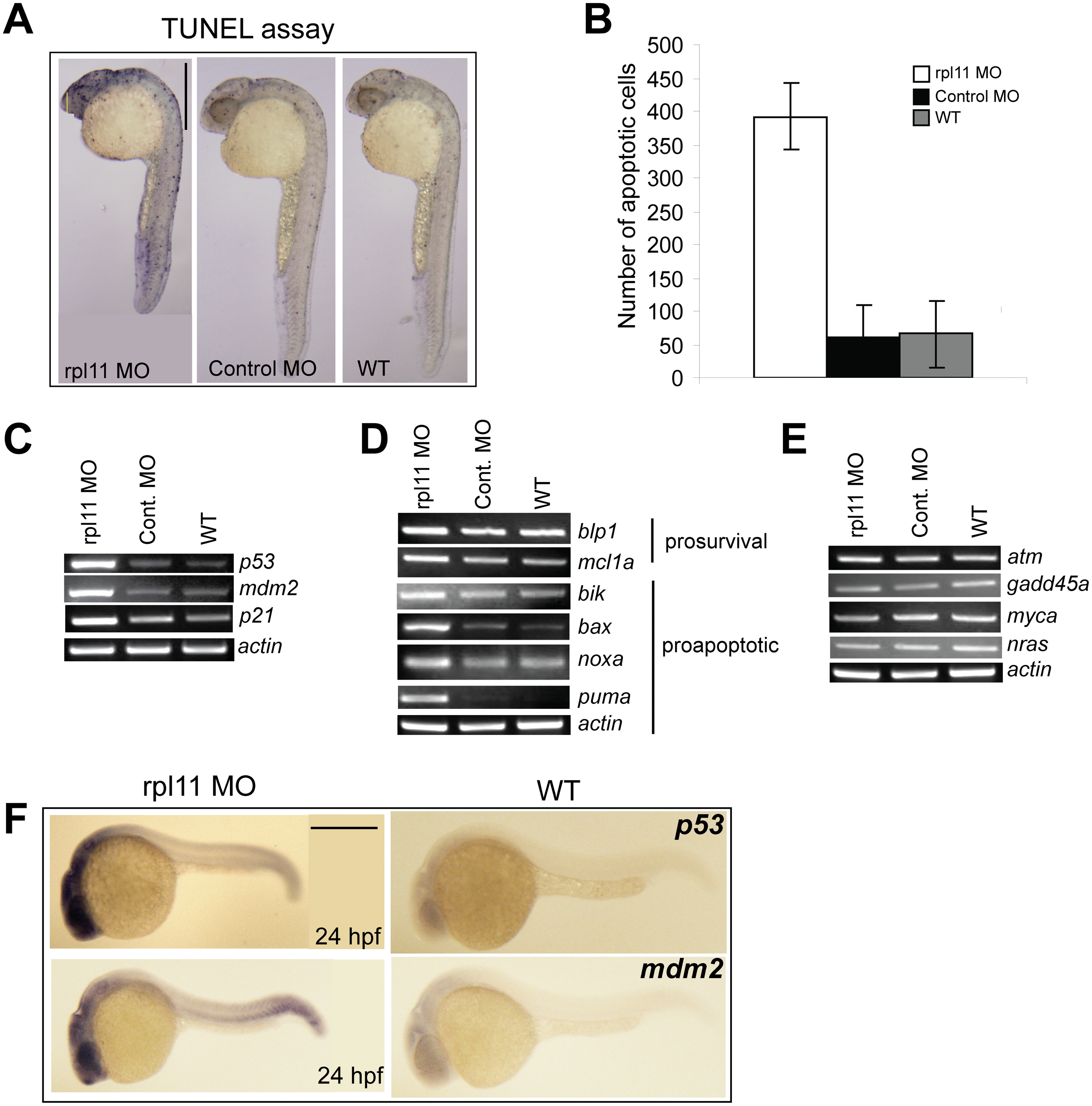
Fig. 4 L11 deficiency results in increased cellular apoptosis mediated by p53.
(A) TUNEL staining at 24 hpf. The morphants show massive apoptosis (blue dots indicate the TUNEL-positive cells) concentrated primarily in the head region compared to a few scattered positive cells in control and wild-type embryos. (B) Histogram showing the number of apoptotic cells in the head region. Values represent the mean of total number of apoptotic cells counted in the head region of three embryos each for the morphant, control, and wild-type embryos. (C-E) Semi-quantitative RT-PCR of p53, p53-target genes, Bcl-2 family genes, atm, gadd45a, myca, and nras transcript levels relative to actin in the morphant, control, and wild-type embryos at 24 hpf. (F) In situ analysis of p53 and mdm2 at 24 hpf. The morphants show increased expression of p53, exclusively in the head region. mdm2 expression, although more ubiquitous, is also increased in the morphants, particularly in the head region. All images are in lateral views with anterior to the top in A and left in F. Scale bars: A, 500 μm.