
Fig. 2
|
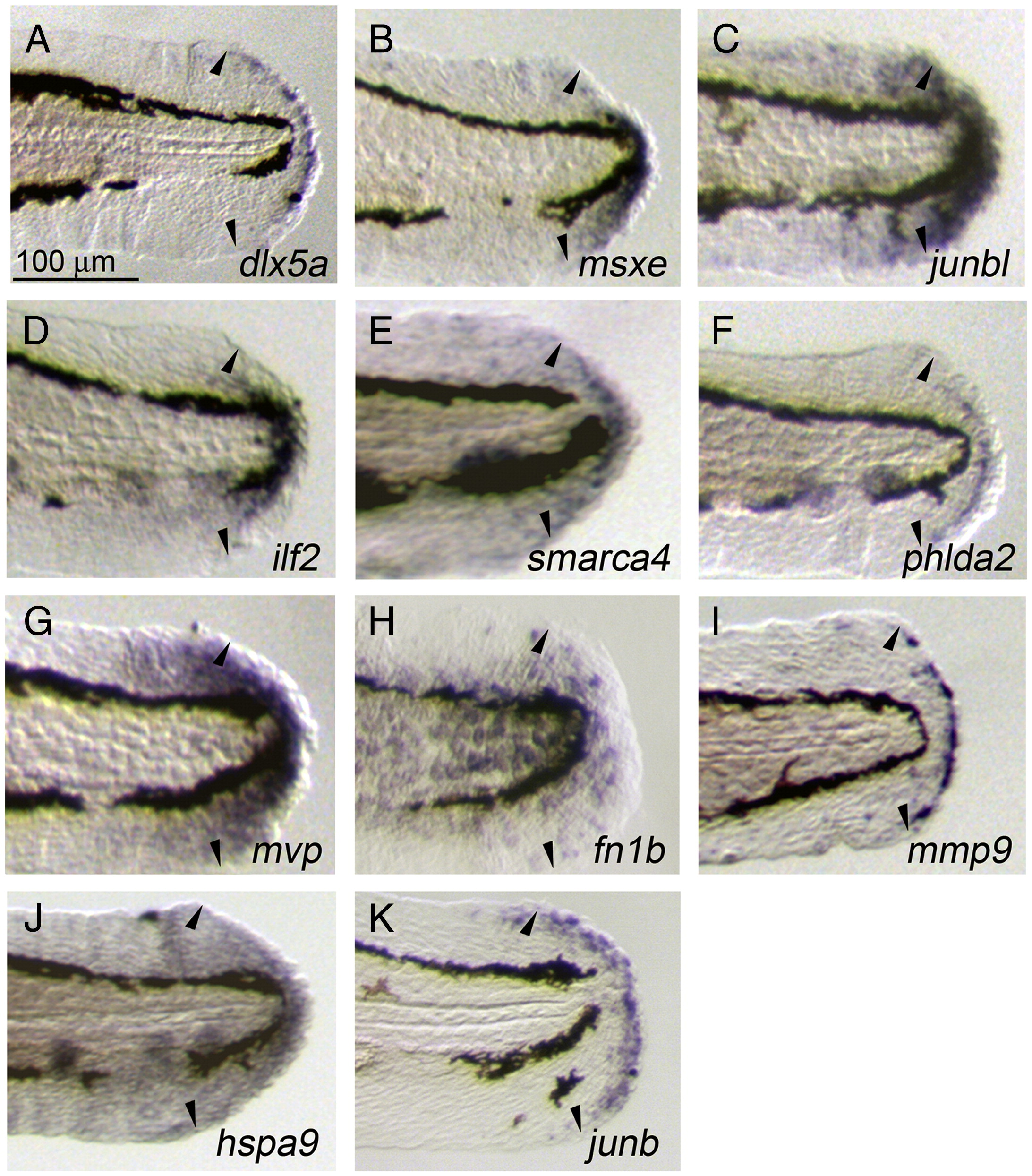
Fig. 2 Induced and localized expression of regeneration-related transcripts during larval regeneration. (A–K) Whole-mount ISH analysis showing the respective gene expression at 1 dpa during larval regeneration. The expressions of dlx5a (A) and junb (K) were seen in the epithelial cells at the stump that appear to correspond to the wound epithelium, whereas the expressions of msxe (B), junbl (C), ilf2 (D), smarca4 (E), mvp (G), and hspa9 (J) were in the mesenchymal regions containing the blastema-like proliferating cells. In addition to these characterized cell populations, the expression of phlda2 was seen in a sharp line of cells between the wound epithelium and mesenchyme (F), mmp9 in a population of epithelial cells with an irregular distribution (I), and fn1b in a broad epithelial region (H). For all genes, expression was not detected in uncut fin folds at the same developmental stage (data not shown). Arrowheads indicate the edges of amputated areas. The same magnifications for panels A–K (scale bar in panel A).
Reprinted from Developmental Biology, 325(1), Yoshinari, N., Ishida, T., Kudo, A., and Kawakami, A., Gene expression and functional analysis of zebrafish larval fin fold regeneration, 71-81, Copyright (2009) with permission from Elsevier. Full text @ Dev. Biol.