
Fig. 6
|
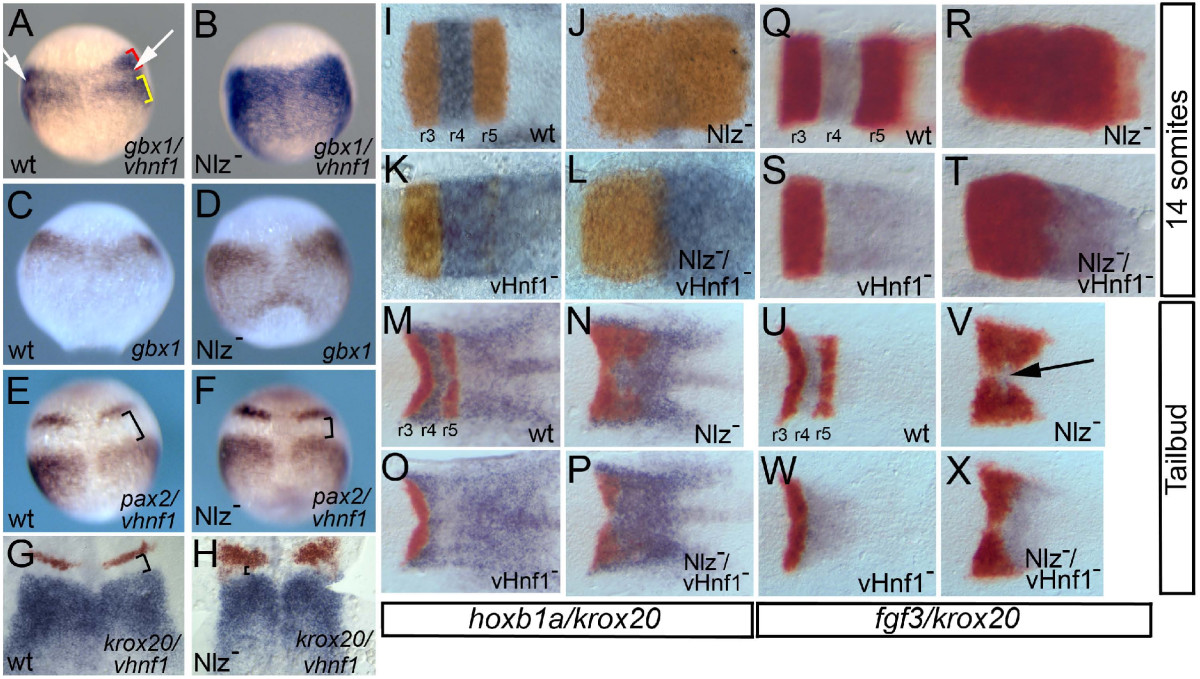
Fig. 6 Nlz proteins act as repressors during zebrafish hindbrain development.A-H. Nlz proteins repress vhnf1 and gbx1 expression in rhombomere 4. Wild type embryos (A, C, E, G) or embryos injected with antisense morpholino oligos targeting Nlz1 and Nlz2 (Nlz-; B, D, F, H) were assayed by whole mount in situ hybridization for expression of gbx1/vhnf1 (A, B: gbx1 red bracket, vhnf1 yellow bracket), gbx1 (C, D), pax2/vhnf1 (E, F; both detected in purple) and krox20/vhnf1 (G, H; krox20 detected in red, vhnf1detected in blue). White arrows in A and black brackets in E-H indicate gaps in gene expression. I-X. Removing vhnf1 function restores late gene expression to Nlz- embryos. Wild type (I, M, Q, U), embryos injected with anti-Nlz MO (Nlz-; J, N, R, V), vhnf1 mutant embryos (K, O, S, W) and vhnf1 mutant embryos injected with anti-Nlz MO (Nlz-/vhnf1-; L, P, T, X) were assayed by in situ hybridization for expression of krox20 /hoxb1a (I-P; krox20 detected in red, hoxb1a detected in blue) or krox20/fgf3 (Q-X; krox20 detected in red, fgf3 detected in blue) at the 14 somite stage (I-L; Q-T) or the tailbud stage (M-P; U-X). Embryos in I-X were dissected and flat-mounted such that only the hindbrain is shown. Anterior is to the top in A-H and to the left in I-X).