
|
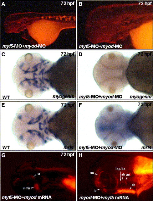
Fig. 5 Myf5 and Myod function independently to activate progenitor lineages in muscles of the head region. Embryos co-injected with 4 ng of myf5-morpholino oligonucleotide (MO) with 4 ng of myod-MO to inhibit specifically both myf5 and myod translation, respectively, were used to observe the development of cranial muscle (A, B) and the expression of myogenin and mrf4 (D, F) at 72 hpf. Panel B was magnified from the head area of panel A. No red fluorescent protein (RFP) signal was detected in muscle primordia in the myf5 and myod double-knockdown morphants derived from the transgenic line Tg(α-actin:RFP; A, B). Similarly, the transcripts of myogenin (C vs. D) and mrf4 (E vs. F) were not detected in the myf5 and myod double-knockdown morphants (I vs. J). The myod mRNA did not rescue the formation of RFP-labeled primordia muscles in myf5 morphants (G). Similarly, the myf5 mRNA did not rescue the formation of RFP-labeled primordia muscle in myod morphants (H). For abbreviations, see the legend of Fig. 1.
Reprinted from Developmental Biology, 299(2), Lin, C.Y., Yung, R.F., Lee, H.C., Chen, W.T., Chen, Y.H., and Tsai, H.J., Myogenic regulatory factors Myf5 and Myod function distinctly during craniofacial myogenesis of zebrafish, 594-608, Copyright (2006) with permission from Elsevier. Full text @ Dev. Biol.