
|
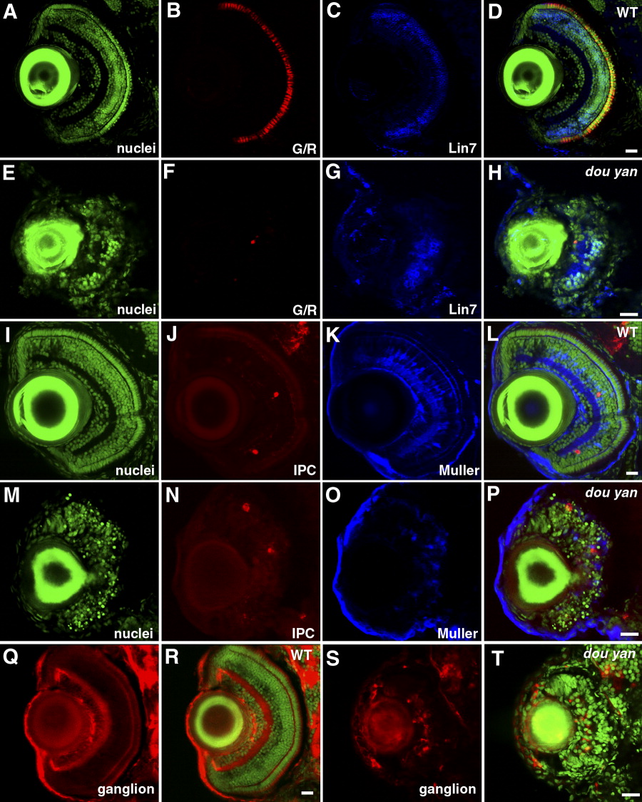
Fig. 7 Distinct types of retinal cells are specified in dou yanmi234 mutants, but they fail to differentiate properly. A-H: At 5 days postfertilization (dpf), green/red double cones (G/R, B and F) and Lin7-positive bipolar cells (Lin7, C and G) are observed in both mutant (E-H) and wild-type (A-D) retinas. YO-PRO staining was used to visualize the entire population of cell nuclei (nuclei, A,E). D and H are merged images. I-P: Interplexiform cells (IPC, J,N) and Muller cells (Muller, K and O) are specified in both wild-type (I-L) and mutant retinas (M-P) at 5 dpf. Cell nuclei were visualized by YO-PRO staining (I,M). L and P are merged images. Q-T: Zn8 (red) -expressing ganglion cells are specified in both wild-type (Q,R) and mutant (S,T) retinas. Cell nuclei were visualized with YO-PRO staining (green). Scale bars = 20 μm.