
Fig. 9
|
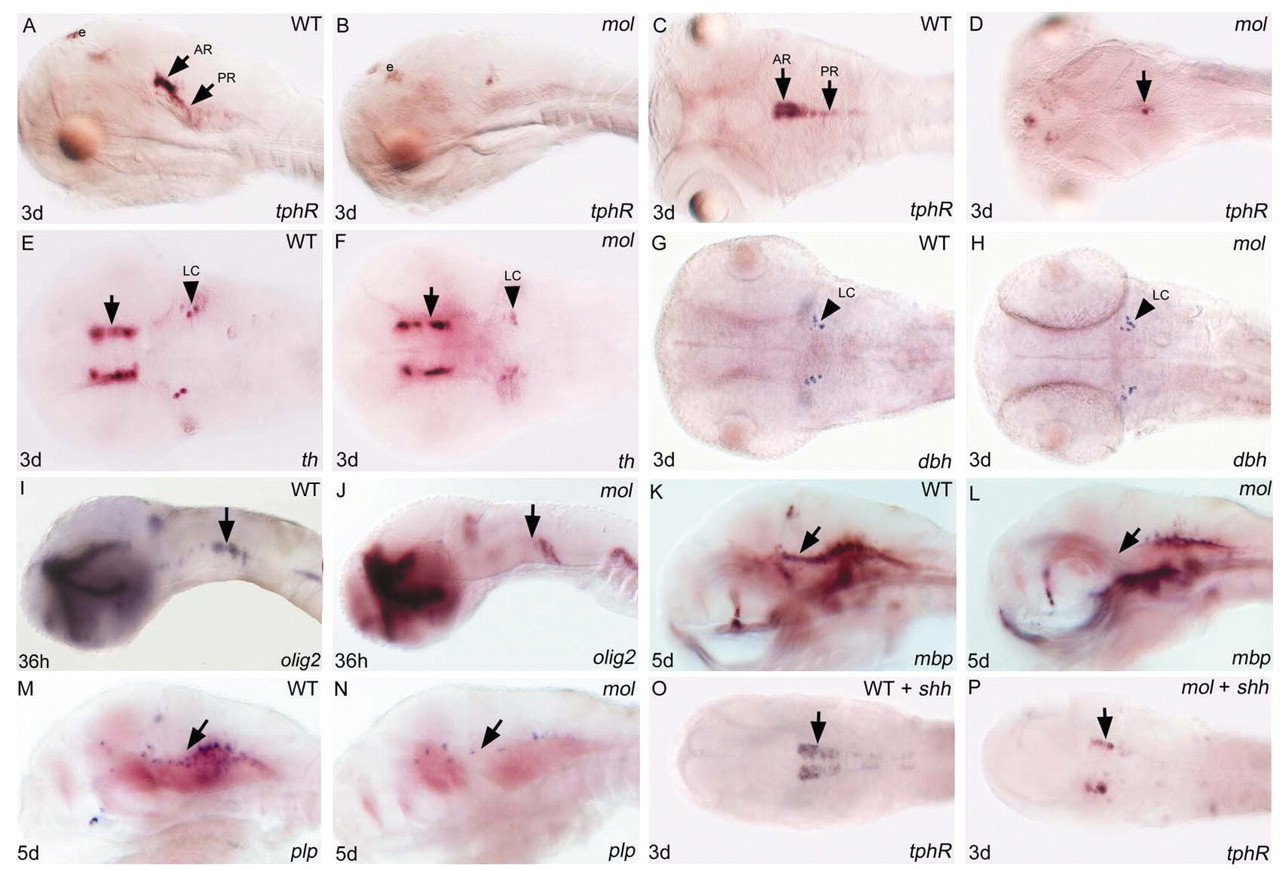
Fig. 9 Both serotonergic raphé neurones and oligodendrocyte precursor cells are severely depleted in mol-/- embryos. Lateral (A,B,I-N) and dorsal (C-H,O,P) views of brains of wild-type and mol-/- embryos and wild-type and mol-/- embryos overexpressing shh (WT + shh; mol + shh) analysed for genes indicated bottom right. (A-D) tphR expression reveals serotonergic neurones. Arrows indicate anterior (AR) and posterior (PR) raphé neurones. Unlabeled arrow in D indicates a few residual tphR + neurons. (E-H) th reveals dopaminergic diencephalic neurones (arrows in E,F); noradrenergic neurones of the LC are labelled both with th and dbh probes. (I-N) olig2 labels oligodendrocyte precursor cells and mbp and plp expression marks nascent oligodendrocytes. Arrows indicate areas of reduced olig2, mbp and plp expression in mol-/- mutant embryos. (O,P) Arrows indicate increased number of tphR-expressing serotonergic neurones in the wild-type embryo and a rescue of serotonergic neurones in the mol-/- embryo following shh injection. e, epiphysis; LC, locus coeruleus.