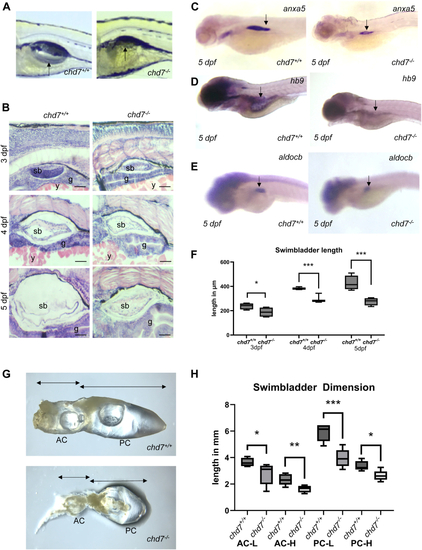
Swim bladder development in chd7−/− mutants. A: Representative images for 3 independent experiments (n = 12 larvae per genotype) of swim bladder development in chd7+/+ and chd7−/− mutants at 5 dpf showing failure to inflate the swim bladder in mutants (arrows). B: HE staining of swim bladder development in chd7+/+ and chd7−/− mutants between 3 and 5 dpf showing generation of the three layers (outer mesothelium, mesenchyme and epithelium). Tissues are labelled g = gut, sb = swim bladder, y = yolk. Scale Bar 50 μm. Representative images for 3 independent experiments (n = 6 larvae per sample/genotype). C: Whole mount in situ hybridization for outer mesothelium marker anxa5 expressed in the swim bladder (arrows) (3 experiments (N = 3), n = 20 larvae for each experiment). D: Whole mount in situ hybridization for epithelium marker hb9 expressed in the swim bladder (arrows) (N = 3, n = 20 larvae for each experiment). E: Whole mount in situ hybridization for mesenchyme marker aldocb expressed in the swim bladder (arrows) (N = 3, n = 20 larvae for each experiment). F: Swim bladder length based on anxa5 WISH (supplemental Fig. S2E) in 3–5 dpf old larvae (N = 3, n = 10 for each experiment). G: Representative adult swim bladder in chd7+/+ and chd7−/− mutants with regions indicated of anterior chamber (AC) and posterior chamber (PC) of the swim bladder. H: Adult swim bladder dimensions, indicating significant smaller anterior and posterior chambers in chd7−/− mutants (chd7+/+ N = 6 and chd7−/− N = 5) (AC: anterior chamber, PC: posterior chamber; H:height; L:length) (Significance: ∗P < 0.05; ∗∗P < 0.01; ∗∗∗P < 0.001, Error bars represent SD).
|
