
FIGURE
Fig. 4
- ID
- ZDB-FIG-251214-27
- Publication
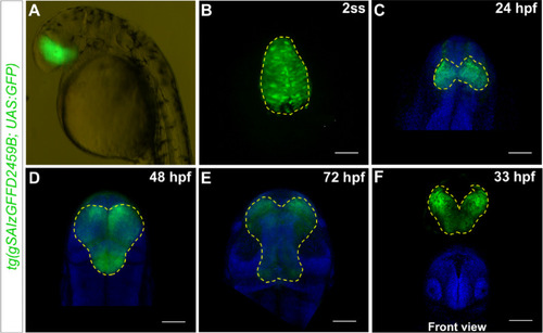
- Vásquez-Ramírez et al., 2025 - A gal4 insertion in the rx3 locus as a tool for visualization and manipulation of eye fated cells in zebrafish
- Other Figures
- All Figure Page
- Back to All Figure Page
Fig. 4
Homozygous |
Expression Data
Expression Detail
Antibody Labeling
Phenotype Data
Phenotype Detail
Acknowledgments
This image is the copyrighted work of the attributed author or publisher, and
ZFIN has permission only to display this image to its users.
Additional permissions should be obtained from the applicable author or publisher of the image.
Full text @ Biol. Res.