|
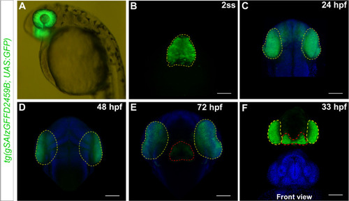
tg(gSAIzGFFD2459B)-driven GFP is expressed in embryonic eyes and diencephalon. A Lateral overview of GFP expression in the eyes of a 36 hpf embryo. B GFP is first visible in the eye field of embryos at the 2-somite stage embryo (dorsal view; anterior is up). C Expression becomes restricted to the optic cups at 24 hpf (dorsal view). D Expression in the eyes is maintained at 48 hpf, while at 72 hpf (E), GFP expression is also observed in the hypothalamus (dorsal view). F A front view at 33 hpf shows the expression pattern in the eyes and hypothalamus. Nuclei are stained with DAPI (blue). Yellow dashed lines outline the GFP expression domains in the eye regions, while red dashed lines mark the hypothalamus of the embryo. Scale bar = 100 µm
|