
Fig. 1
- ID
- ZDB-FIG-251107-5
- Publication
- Ramírez et al., 2025 - The Smarce1 subunit of the BAF complex performs distinct, stage-specific functions during zebrafish retinal development
- Other Figures
- All Figure Page
- Back to All Figure Page
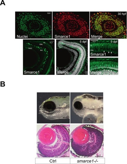
Immunodetection of Smarce1 in retinas at 30 hpf and 5 dpf. (A) Confocal sections of a 30 hpf retina stained with a Smarce1 antibody (green) (upper panel); the signal is detected in all RPCs nuclei. Cryostat histological sections of 5 dpf retinas showing DAPI stained nuclei (gray) and Smarce1 signal (green) in amacrine cells (Am), retinal ganglion cells (RGC), Photoreceptor layer (PL), and outer nuclear layer (ONL) (lower panel). The high magnification images on the right show Smarce1 signal in most photoreceptor nuclei, except for a few basally-localized ones, which appear negative (arrowheads). Signal in outer segments (PL) is due to autofluorescence. (B) Bright-field photographs of wild type (Ctrl) and mutant (smarce1-/-) heads showing the eye size reduction (upper panel). H&E sections of 5 dpf wild type (Ctrl) and mutant (smarce1-/-) retinas (lower panel). Scale bars in A: 20 μm; in B: 50 μm. (For interpretation of the references to colour in this figure legend, the reader is referred to the web version of this article.) |