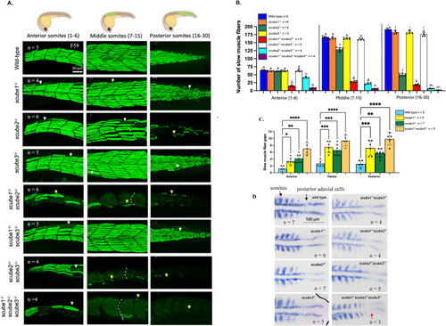
scube mutants display Hh loss-of-function phenotypes. (A) Loss of slow muscle fibres in scube mutant families at 30 hpf. In wild-type zebrafish, slow muscle fibres form a regular array with clearly defined V-shaped somites. In scube1−/−, scube3−/−, and scube1−/−scube3−/−, morphology of slow muscle fibres are comparable to wild-type siblings. In contrast, scube2−/−, scube1−/−scube2−/−, scube2−/−scube3−/−, and the triple scube mutant show severe disruption of slow muscle fibres along the body, including fibre loss (white arrows), abnormal fibre shapes (yellow arrows), and the presence of U-shaped somites (indicated by white dashed lines). The triple mutant shows the most severe phenotype, characterised by the near-complete loss of slow muscle fibres and pronounced abnormalities in the remaining fibres. (B) Slow muscle fibres were quantified in the anterior (somites 1–6), middle (somites 7–15), and posterior (somites 16–30) regions of 30 hpf embryos. In each region, bars labelled with different letters (a–m) represent statistically significant differences. For example, in anterior somites, the number of slow muscle fibres in wild-type, scube1−/−, scube3−/−, and scube1−/−scube3−/− were labelled ‘a' because they are not statistically different from each other; however, they are statistically different from scube1−/−scube2−/− (labelled ‘b'), scube2−/−scube3−/− (labelled ‘c'), and triple scube knockout (labelled ‘d'). Statistical analysis was performed using ANOVA with multiple comparisons and Bonferroni correction. The numbers of slow muscle fibres in scube1−/−, scube3−/−, and double scube1−/−scube3−/− mutants are not different from the wild-type, except in the posterior region of the double mutant, where fibre number is significantly reduced compared to wild-type siblings. Combining scube2 KO with either scube1 or scube3 KO results in a significantly greater reduction in muscle fibres than in single mutants. The triple mutant has the lowest number of slow muscle fibres among all genotypes tested. (C) Fibre gaps in scube1−/−, scube3−/−, and scube1−/−scube3−/− mutants at 30 hpf were counted. Each fibre gap comprised approximately one to four fibres. Statistical analysis showed a significant increase in the number of fibre gaps in each mutant compared to wild-type siblings. Statistical analysis was performed using ANOVA with multiple comparisons and Bonferroni correction. (D) myoD staining in the scube mutant family at 15-somite stage, n = 3 to 7 per mutant. Dorsal views with the head oriented to the left. In wild-type, single, and double scube mutants, myoD expression is expressed in adaxial cells and somites. In the triple mutant (scube1−/−scube2−/−scube3−/−), myoD expression in posterior adaxial cells is lost (red arrow), indicating severe disruption of Hh signalling.
|
