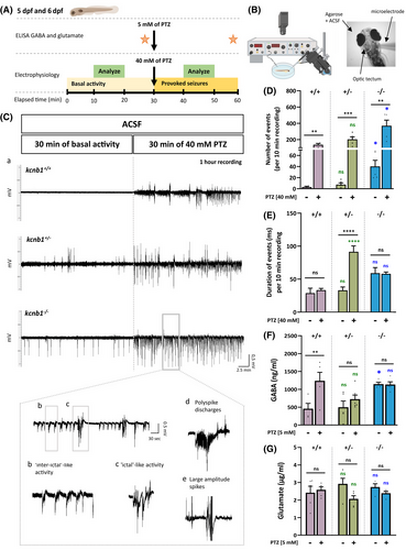
kcnb1−/− larvae show spontaneous and provoked epileptiform-like electrographic activity associated with a dysregulation of γ-aminobutyric acid (GABA). (A, B) Schematic representation of the protocol applied for electroencephalographic recordings and neurotransmitter quantification of 5 and 6 days post-fertilization (dpf) zebrafish. Neuronal activity in the optic tectum of zebrafish was recorded by applying a protocol of 30 min of basal activity followed by 30 min of 40mmol·L−1 pentylenetetrazol (PTZ) treatment. A 10-min segment in the middle of each period (basal activity and provoked seizures) was used to analyze the total number of negative spikes and duration of events for each genotype. Enzyme-linked immunosorbent assay (ELISA) was performed to quantify GABA and glutamate following a 30-min pre- and post-PTZ treatment at 5 mmol·L−1. (C; a) Representative traces of electroencephalographic recordings in the optic tectum of kcnb1+/+ and kcnb1 knockout (kcnb1+/− and kcnb1−/−) larvae showing spontaneous and provoked epileptiform-like electrographic activity in the kcnb1−/− model characterized by (b) interictal-like activity, (c) ictal-like activity, (d) polyspike discharges, and (e) large-amplitude spikes. (D, E) Quantification of electrophysiological recordings by analyzing (D) the total number of negative spikes and (E) the duration of events, over 10 min as described in panel A. kcnb1−/− larvae showed significantly increased spontaneous and provoked neuronal activity, reflected by a significant elevation of the number of spikes, although the duration of events was similar to the kcnb1+/+ condition in pre- and post-PTZ treatment. kcnb1+/− larvae presented a profile similar to the kcnb1+/+ condition in terms of number of spikes but showed a significant increase of event duration (see Table S3; n = 3–5/genotype; unpaired t-test). (F, G) Quantification of (F) GABA and (G) glutamate by ELISAs in the head of 6-dpf larvae following the 30-min pre- and post-5mmol·L−1 PTZ treatment. kcnb1−/− zebrafish present significantly increased GABA levels observed in both baseline and post-PTZ conditions conversely to kcnb1+/− larvae and compared to the kcnb1+/+ condition. We observed a lack of significant changes in glutamate levels in both kcnb1 knockout models (kcnb1+/− and kcnb1−/−) in pre- and post-PTZ conditions (N = 3–5; n = 50/sample; unpaired t-test). For panels D–G, colored statistic indications correspond to mutant conditions compared to the kcnb1+/+ condition in pre- or post-PTZ treatment. *p < .05, **p < .01, ***p < .001, ****p < .0001. ACSF, artificial cerebrospinal fluid; ns, non-significant.
|
