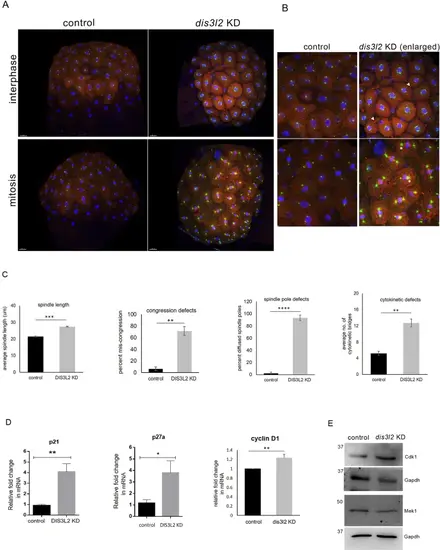
dis3l2 is required for early embryonic mitoses A: Sum projection confocal image of 256 cell stage control and dis3l2 morphants embryos showing mitotic defects. Blue/DAPI - DNA, red– alpha-tubulin, green– γ tubulin. B: Enlarged sum projection confocal image of dis3l2 morphants showing cytokinetic index (white arrowhead), spindle length (white dotted line), and spindle poles (white asterisks) during early mitoses. Scale bars, 50 μm. C: Quantification of spindle length (white dotted line), chromosome congression, spindle pole (white asterisk), and cytokinesis (white arrowhead) in the dis3l2 morphants as compared to control. Scale bars, 50 μm. All data are shown as mean + SEM. *p < 0.05, **p < 0.01, ***p < 0.001. n = 3 for each experiment, with a minimum of 20 embryos per experiment. D: Relative expression of cell cycle regulators– p21, p27, and cyclinD1 in dis3l2 morphants. Data are shown as mean + SEM, *p < 0.05, **p < 0.01, ***p < 0.001. n = 3 for each experiment, with a minimum of 100 embryos per experiment. E: Western blot analysis showing Cdk1 and Mek1 levels in dis3l2-depleted embryos as compared to control. Gapdh was used as the loading control
|
