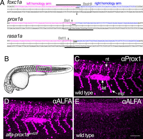
Targeted insertion of an ALFA tag using optimized knock-in parameters. (A) Sequence and flanking target insertion sites for foxc1a, prox1a and rasa1a. Left and right homology arms are indicated by magenta and blue, respectively. Black lines denote the Cas9 spacer sequence (line above or below sequence denotes positive or negative strand, respectively) and arrowheads mark the DSB sites. Start or stop codons are underlined. Restriction enzyme sites used to estimate nuclease activity are indicated by boxes and labeled. (B) Camera lucida of a zebrafish embryo at 31 hpf. Reproduced with permission from Kimmel et al. (1995). (C-E) Whole-mount immunostained embryos imaged by confocal microscopy; lateral views, anterior to the left, dorsal is up. (C) Wild-type embryos immunostained with polyclonal Prox1 antibody. Arrows denote fluorescence in neural tube (nt), lateral line primordium (lat) and muscle pioneers (mp). (D,E) Immunostaining of alfa-prox1aum529 heterozygous (D) and wild-type sibling (E) embryos with a nanobody against ALFA. Scale bar: 80 µm.
|
