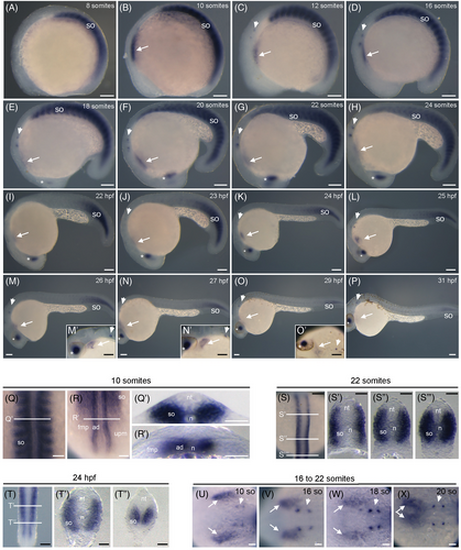
Spatio-temporal expression of rbm24a transcripts during zebrafish development. (A–P) Lateral views, with anterior to the left, showing rbm24a expression in the developing somites from 8- to 24-somite stages (A–H) and progressively restricted to most caudal somites from 22 hpf to 31 hpf (I–P). (M′—O′) Higher magnifications of the anterior region. (Q, R) Dorsal views, with anterior to the top, of the embryo at 10-somite stage shown in B, at the level of the anterior and posterior re-gions, respectively. (Q', R') Transverse sections at the levels indicated in Q and R, respectively. Note that rbm24a is expressed in the entire somite and in adaxial cells of the presomitic mesoderm. (S) Dorsal view, with anterior to the top, of the embryo at 22-somite stage shown in G, focused on the posterior region. (S′–S″′) Transverse sections at the level indicated in S. (T) Dorsal view, with anterior to the top, of the embryo at 24 hpf shown in K, focused on the most posterior region. (T′, T″) Transverse sections at the level indicated in T. (U–X) Dorsal views, with anterior to the right, of embryos presented in B–G, at the level of the heart area. The expression of rbm24a is also present in the developing heart (white arrows), otic placode (arrowheads), and lens (asterisks). So, somite; nt, neural tube; n, notochord; ad, adaxial cells; fmp, fast muscle progenitors; upm, unsegmented presomitic mesoderm. Scale bars: 100 μm.
|
