|
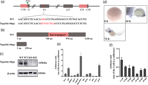
Targeted disruption of the zebrafish trpa1b gene using clustered regularly interspaced short palindromic repeats (CRISPR)/Cas9. (a) Structure of the zebrafish trpa1b gene and target site. The mutant site contains a − 3 bp (ATG) DNA sequence and a + 9 bp (TCCGCTGAC) insertion sequence, which includes a stop codon. (b) Schematic representation of the wild-type (WT) and Trpa1b proteins. Functional domains are indicated in red and consist of amino acids (aa) from 760 to 976, which do not reach the ion channel domain. (c) Detection of Trpa1b protein expression at 60 h postfertilization (hpf). KO: Trpa1b−/−. (d) Whole-embryo in situ hybridization showing the temporal expression of trpa1b. (e–f) Analyse the relative expression levels of trpa1b messenger RNA (mRNA) in different adult tissues and embryonic development stages using real-time quantitative PCR (RT-qPCR). The data are presented as mean ± SE of mean (n = 3, biological replicates).
|