|
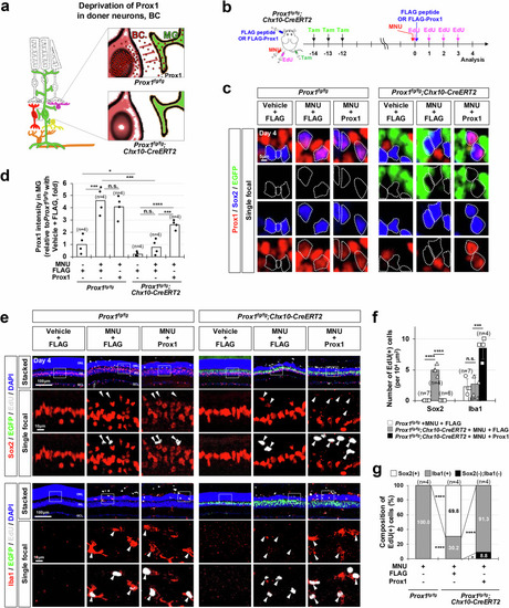
Prox1 gene deletion in donor retinal neurons restores MG proliferative potential. a Schematic representation of Prox1 depletion in MG achieved through Prox1 gene deletion in BC. (b) Prox1 was selectively deleted and EGFP was complementarily expressed in BCs of Prox1fg/fg;Chx10-CreERT2 mouse retinas by repeated Tamoxifen (Tam) injections. Following this, mice were injected with MNU to induce PR degeneration and EdU to label proliferating cells. As indicated, FLAG-Prox1 recombinant protein (250 fmol) or FLAG peptides were injected intravitreally. c Distribution of Prox1, Sox2, and EGFP in the retinas of Prox1fg/fg and Prox1fg/fg;Chx10-CreERT2 littermates was assessed by immunostaining. Sox2-positive MG nuclei are outlined by dotted-lines. d Relative Prox1 immunofluorescent intensity in MG in the corresponding retina, normalized to Prox1 intensity in BCs within the same image, is shown. Each dot represents the median intensity collected from one retina. Number of samples analyzed is 4. e MG and microglial identities of EdU-labeled newborn cells in mouse retinas were determined by co-staining Sox2 and Iba1. The boxed areas in the top row are enlarged in the following two rows. Arrowheads point to EdU-labeled cell nuclei. f Quantification of EdU-labeled MG and microglia in the retinas is shown in the graph. g Composition of EdU-labeled cells in the mouse retinas is displayed in the graph. Numbers of samples analyzed are shown in the graph (data from 4 independent litters). Error bars denote SEM. P-values were calculated using one-sided Student’s t-test (***, p < 0.005; ****, p < 0.001; n.s., > 0.05).
|