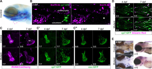
Normal patterns of growth and differentiation in sox10 mutant osteoblasts. (A) Reference image of a larva stained with Alcian Blue and Alizarin Red, with locations of the skeletal elements shown in B (magenta dashed box) and C (white box) highlighted. (B) Immunostaining with an anti-Sox10 antibody reveals strong expression in chondrocytes but a lack of Sox10 protein (asterisk) in mineralizing osteoblasts (sp7:GFP+) forming the op bone at 3 dpf. (C-C″) Example images from sequential live imaging showing normal patterns of RUNX2:mCherry, sp7:GFP and osc:GFP transgene expression in mutant osteoblasts of the op at 4 and 7 dpf. (D) Normal growth of sox10 mutant op (arrowheads) as well as other bones despite minimal calcium accumulation, revealed by live imaging of Alizarin Red-stained sp7:GFP+ embryos at 4 and 7 dpf. (E) Colorimetric in situ hybridizations for col10a1a and phospho1, encoding key bone matrix components, revealed no overt abnormalities in sox10 mutants at 4 dpf. op, opercle. Numbers in panels indicate the proportion of larvae of that genotype with the presented phenotype. Scale bars: 10 µm (B-C″); 100 µm (D,E).
|
