
Fig. 5
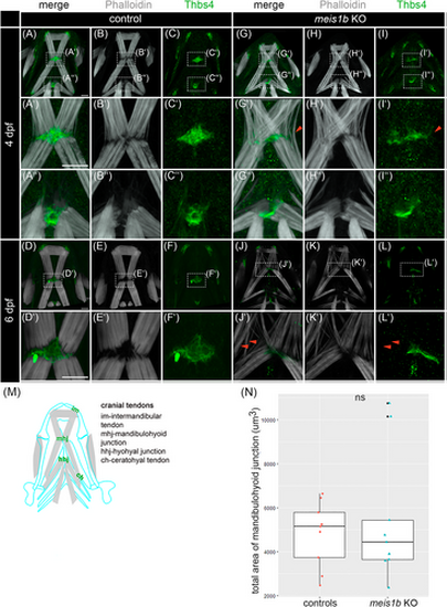
Tendons are disorganized together with muscles in meis1b mutants. (A–L′) Immunostaining of tendons with Thbs4 antibody (green) and muscles with phalloidin (white). Ventral views with the anterior to the top. (A′–C′, G′–I′) Detail of the mandibulohyoid junction at 4 dpf. (A″–C″, G″–I″) Detail of the hyohyal junction at 4 dpf. (G′, I′) Red arrowheads indicate affected muscle fibers without tendon. (D′–F′, J′–L′) Detail of the mandibulohyoid junction at 6 dpf. (J′, L′) Tendons and muscles are more disorganized, and some muscle fibers lacked corresponding tendons (red arrowheads). (M) Schematic position of tendons in the context of head muscles (gray) and cartilage (blue contours) at 6 dpf. (N) Quantification of the total area of the mandibulohyoid junction, color dots indicate individual animals (controls = 9, mutants = 9). Mann–Whitney U test was used for statistical analysis. Scale bar = 30 μm. |