
Fig. 7
- ID
- ZDB-FIG-240904-26
- Publication
- Ma et al., 2024 - Instantaneous visual genotyping and facile site-specific transgenesis via CRISPR-Cas9 and phiC31 integrase
- Other Figures
- All Figure Page
- Back to All Figure Page
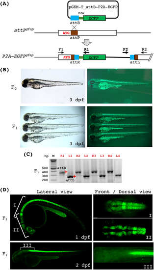
Generation of the endogenous gfap reporter line via phiC31 integrase. (A) A diagram depicting the integration of a promoter-less EGFP reporter at the attPgfap locus via phiC31-mediated recombination. Correct integration of pGEM-T_attB-P2A-EGFP will result in fusion and co-translation of the N-terminus of gfap and P2A-EGFP. We named this allele P2A-EGFPgfap. Two sets of PCR primers (F1/R1 and F2/R2 as indicated) were used to detect the integration. F1 and R2 are gfap-specific, whereas R1 and F2 are plasmid-specific. (B) Bright-field and fluorescence images of endogenous gfap reporter fish embryos at 3 dpf. The top panels show an attPgfap embryo after microinjection with the phiC31 mRNA and pGEM-T_attB-P2A-EGFP. The bottom panels show three heterozygous P2A-EGFPgfap embryos. (C) PCR analysis of individual fluorescent embryos from a founder fish indicating that all of them harbored the correct integration. ‘R’ and ‘L’ in front of the numbers on top indicate attR and attLPCR, respectively. The red shining star symbols mark the correct size products of attR (332 bps) and attL (282 bps). (D) Stacked confocal images of the P2A-EGFPgfap embryos showing various regions and angles of view at 1 and 2 dpf. |