FIGURE

Fig. 3

ID
ZDB-FIG-240118-27
Publication
Voigt et al., 2023 - Reflective multi-immersion microscope objectives inspired by the Schmidt telescope
Other Figures
All Figure Page
Back to All Figure Page
Fig. 3

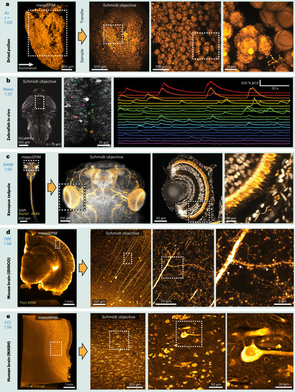
Example two-photon datasets acquired with the Schmidt objective.

Fixed samples were first imaged with a mesoSPIM light-sheet microscope before the sample was transferred to the Schmidt objective. a, Pollen pellet composed of many individual pollen grains imaged in air. b, Functional imaging in a 5-day-old elavl3:GCaMP6s zebrafish larva in vivo (example dataset from one of three imaged larvae). c, BABB-cleared X. tropicalis tadpole stained for Atp1a1 (Alexa Fluor 594, orange) and nuclei (DAPI, grayscale). The large Schmidt FOV allows both imaging of the entire head (≈800 µm across) and imaging of individual developing photoreceptors in the eye (right). The images are examples from one of four imaged tadpoles. d, Thy1-H-labeled coronal mouse brain slice imaged in DBE. At higher magnification, spines on apical dendrites of L5 neurons are visible. The images are examples from one of two imaged mouse brain slices. e, MASH-processed human neocortex stained with acridine orange. The images are examples from one of two imaged samples.

Expression Data

Expression Detail
Antibody Labeling
Phenotype Data

Phenotype Detail
Acknowledgments
This image is the copyrighted work of the attributed author or publisher, and ZFIN has permission only to display this image to its users. Additional permissions should be obtained from the applicable author or publisher of the image. Full text @ Nat Biotechnol.