FIGURE SUMMARY
Title

Reflective multi-immersion microscope objectives inspired by the Schmidt telescope

Authors
Voigt, F.F., Reuss, A.M., Naert, T., Hildebrand, S., Schaettin, M., Hotz, A.L., Whitehead, L., Bahl, A., Neuhauss, S.C.F., Roebroeck, A., Stoeckli, E.T., Lienkamp, S.S., Aguzzi, A., Helmchen, F.
Source
Full text @ Nat Biotechnol.

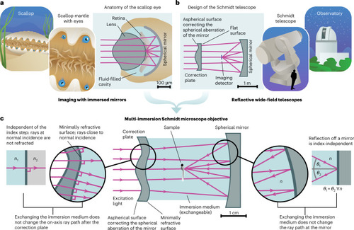
Concept of the multi-immersion Schmidt objective.

a, Our approach is inspired by the anatomy of the scallop eye. In this eye design, a curved mirror in contact with a liquid forms an image on a transparent layer of photoreceptors. b, A second inspiration is the Schmidt telescope, a mirror-based wide-field telescope design that consists of a spherical mirror and an aspherical correction plate. c, We synthesize both concepts into a multi-immersion objective design that provides a sharp image in any homogeneous medium (center). Reflection off the mirror does not depend on the refractive index n of the immersion medium (right). To avoid additional refraction at the inner surface of the correction plate, we deform this surface such that all passing rays are close to normal incidence (left).

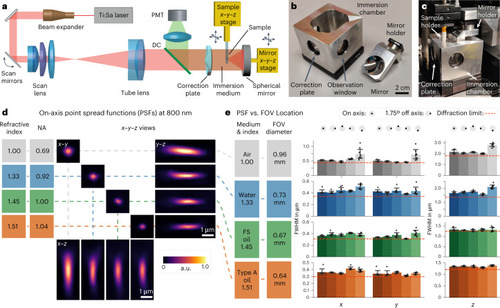
Setup and characterization.

a, Overview of the multi-photon microscopy setup. In our prototype, the position of the spherical mirror needs to be aligned relative to the correction plate for optimum image quality. This design allows for straightforward cleaning of the mirror when switching immersion media. To create a z-stack, the sample is moved along the optical axis. DC indicates a dichroic mirror that separates the emitted fluorescence from the excitation beam. b, The prototype objective consists of an immersion chamber and a mirror. c, Assembled microscope objective. d, xyz views of PSF measurements of 200-nm fluorescent beads imaged at refractive indices ranging from n = 1.00 (air), n = 1.33 (water) and n = 1.45 (FS, Cargille fused silica matching oil) to n = 1.51 (Cargille oil type A). Because NA is proportional to n, an increase in n leads to higher NA and, thus, a smaller PSF. e, PSF uniformity over the FOV. PSF measurements were carried out on-axis and at a scan angle of 1.75°, which was simulated to be the maximum theoretical diffraction-limited scan angle for the total system (Extended Data Fig. 1). As the magnification of the objective is proportional to n, the FOV diameter decreases with increasing index. Diffraction-limited theoretical values are indicated by the red dashed lines. The PSF FWHM (±s.e.m.) was measured for eight beads for each FOV location and immersion medium. a.u., arbitrary units.

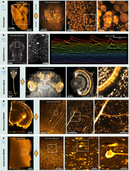
Example two-photon datasets acquired with the Schmidt objective.

Fixed samples were first imaged with a mesoSPIM light-sheet microscope before the sample was transferred to the Schmidt objective. a, Pollen pellet composed of many individual pollen grains imaged in air. b, Functional imaging in a 5-day-old elavl3:GCaMP6s zebrafish larva in vivo (example dataset from one of three imaged larvae). c, BABB-cleared X. tropicalis tadpole stained for Atp1a1 (Alexa Fluor 594, orange) and nuclei (DAPI, grayscale). The large Schmidt FOV allows both imaging of the entire head (≈800 µm across) and imaging of individual developing photoreceptors in the eye (right). The images are examples from one of four imaged tadpoles. d, Thy1-H-labeled coronal mouse brain slice imaged in DBE. At higher magnification, spines on apical dendrites of L5 neurons are visible. The images are examples from one of two imaged mouse brain slices. e, MASH-processed human neocortex stained with acridine orange. The images are examples from one of two imaged samples.

Acknowledgments
This image is the copyrighted work of the attributed author or publisher, and ZFIN has permission only to display this image to its users. Additional permissions should be obtained from the applicable author or publisher of the image. Full text @ Nat Biotechnol.