|
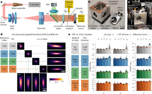
Setup and characterization. a, Overview of the multi-photon microscopy setup. In our prototype, the position of the spherical mirror needs to be aligned relative to the correction plate for optimum image quality. This design allows for straightforward cleaning of the mirror when switching immersion media. To create a z-stack, the sample is moved along the optical axis. DC indicates a dichroic mirror that separates the emitted fluorescence from the excitation beam. b, The prototype objective consists of an immersion chamber and a mirror. c, Assembled microscope objective. d, x–y–z views of PSF measurements of 200-nm fluorescent beads imaged at refractive indices ranging from n = 1.00 (air), n = 1.33 (water) and n = 1.45 (FS, Cargille fused silica matching oil) to n = 1.51 (Cargille oil type A). Because NA is proportional to n, an increase in n leads to higher NA and, thus, a smaller PSF. e, PSF uniformity over the FOV. PSF measurements were carried out on-axis and at a scan angle of 1.75°, which was simulated to be the maximum theoretical diffraction-limited scan angle for the total system (Extended Data Fig. 1). As the magnification of the objective is proportional to n, the FOV diameter decreases with increasing index. Diffraction-limited theoretical values are indicated by the red dashed lines. The PSF FWHM (±s.e.m.) was measured for eight beads for each FOV location and immersion medium. a.u., arbitrary units.
|