|
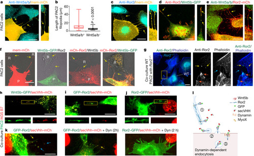
Wnt5b–Ror2 complexes are transported from the producing cells to the receiving cells via cytonemes. a,c–e, PAC2 cells transfected with mem-mCherry (a,c), Wnt5b–GFP (d) or Ror2–mCherry (e) and stained with an antibody against Wnt5a/b (a,e) or against Ror2 (c,d). Yellow arrows, Wnt5a/b–Ror2 on cytonemes. mCh, mCherry. Scale bars 10 µm. b, Length of filopodia with or without Wnt5a/b (n = 17 cells, 3 biological repeats). Two-tailed P values by Mann–Whitney test. Box and whisker plots show median, and top and bottom quartile ranges, with whiskers extending to minimum and maximum values. f, PAC2 cells were transfected with indicated markers and imaged live at 24 h post-transfection. Yellow arrows, Wnt5b (second from left) Ror2 (third from left) and Wnt5b–Ror2 (right) in the non-transfected neighbouring cells. R, receiving cells; P, producing cells. Scale bar, 5 µm. Left to right: n = 36, 38, 42 and 28 cells, 3 biological repeats. g, Left, wild-type (WT) PAC2 cells were co-cultivated with Ror2–/– cells for 24 h, then stained with a Ror2 antibody. The area bound in yellow is magnified in the other images. Orange arrows, Ror2 puncta at the plasma membrane; yellow arrows, Ror2 puncta in the adjacent Ror2−/− PAC2 cells; blue arrows, filopodia. Scale bar, 10 µm. n = 46 cells, 3 biological repeats. h–k, PAC2 cells transfected with indicated constructs, co-cultured with AGS cells expressing secVhh–mCherry, PAC2 cells transfected with Wnt5b–GFP (h), GFP–Ror2 (i,k), Ror2–GFP (j) and treated with 40 µM dynasore (Dyn) (k). h–j, The region bound in yellow shows protrusions, and is magnified below the main image. n = 24 (h), 22 (i), 36 (j) cells, 3 biological repeats. k, Co-localization on cytonemes (yellow arrows) and in receiving cells (blue arrows). Left to right: n = 22, 38 and 9 cells, 3 biological repeats. Scale bars, 10 µm. l, Schematic of the handover mechanism: (1) transport along cytonemes; (2) deposition of cargo-positive vesicles; and (3) endocytosed vesicles. Source Data
|