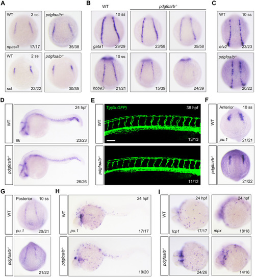
pdgfαa and pdgfαb are necessary for erythroid progenitor differentiation but are dispensable for the development of endothelial and myeloid lineages. (A) Expression patterns of npas4 l and scl in wild-type (WT) and pdgfαa−/−;pdgfαb−/− (pdgfαa/b−/−) embryos at the two-somite stage (2 ss) as revealed by in situ hybridization. Dorsal view. (B) Expression of gata1 and hbbe3 in WT and pdgfαa−/−;pdgfαb−/− embryos at the 10-somite stage (10 ss) assayed by in situ hybridization. Dorsal view. (C,D) In situ hybridization expression analysis of (C) etv2 (dorsal view) and (D) flk (lateral view with head to the left) in WT and pdgfαa−/−;pdgfαb−/− embryos at the indicated stages. (E) Confocal images of pdgfαa−/−;pdgfαb−/− mutants in the Tg(flk:GFP) background at 36 hpf. Lateral view. Scale bar: 50 μm. (F-H) Expression patterns of pu.1 in WT and pdgfαa−/−;pdgfαb−/− embryos at the indicated stages as revealed by in situ hybridization. Dorsal view. (I) Expression analysis of lcp1 and mpx in WT and pdgfαa−/−;pdgfαb−/− embryos at 24 hpf by in situ hybridization. Ventral view in the left panel and lateral view in the right panel. Images shown are representative of the observed phenotypes. The number of embryos displaying each phenotype out of the total number assayed is indicated.
|
