|
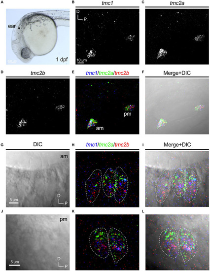
Overlapping expression of tmc1/2a/2b in nascent hair cells at 1 dpf. (A) Image of the anterior region of a 1 dpf embryo. Otocyst indicated by asterisk. Expression of tmc1(B), tmc2a(C), and tmc2b(D). Merge of all three channels (E) and overlay with a DIC image (F). In merged images, fluorescent label for tmc1 transcripts is shown in blue, tmc2a in green, and tmc2b in red. (G–L) Single optical sections (0.75 μm) of overlapping expression of tmc1/2a/2b in hair cells at 1 dpf. Dotted outlines indicate the somas of single hair cells. (G–I) Lateral view of three hair cells in the anterior macula at 1 dpf with visible apical structures. (J–L) Lateral view of the cell bodies of two hair cells in the posterior macula at 1 dpf (kinocilia are not present within the same section).
|