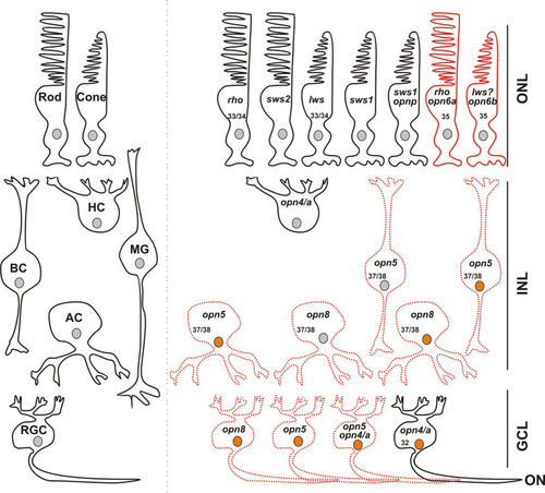
Opsin-expressing cells in the retina of a stage 43/44 Xenopus tadpole. Photoreceptors located in the outer nuclear layer (ONL) express the classical opsins in rods (rho or sws2; Kojima et al., 2017) and cones (lws and sws1). Pinopsin (opnp) is co-expressed in a few sws1+ cells (Bertolesi et al., 2021). Labeled with red are cells expressing opn6a and opn6b in rods (rho+) and cones, likely lws+, respectively. In the inner nuclear layer (INL), horizontal cells (HC) co-express the two melanopsin genes (opn4 and opn4a), bipolar cells (BC) express opn5, while amacrine cells (AC) express opn5 or opn8. Different cells in the ganglion cell layer (GCL) express opsins, including opn5, opn8, opn4/opn4a, and a few cells that contain opn4/a and opn5. Cells that express c-fos in response to exposure to white light are shown with an orange nucleus. Although classical photoreceptors are c-fos negative, they are highly likely activated by the white light exposure. We propose that opsin-expressing cells in the INL or GCL that exhibit light-induced c-fos expression serve as second- or third-order neurons, which receive synaptic inputs from photoreceptors or other retinal cells. We cannot rule out, however, that the opsin-expressing cells of the INL and GCL are intrinsically photosensitive and induce c-fos mRNA upon light stimulation, acting as first-order neurons. opn4/a+ HCs do not express c-fos while opn4/a+ RGCs do (Bertolesi et al., 2014). Opsin-expressing cells that we describe in this manuscript are either labeled with red solid lines (PR; photoreceptors), or red dotted lines as having the potential to be photosensitive in Xenopus, since direct light activation has not yet been demonstrated. Excluded are all the photoreceptors, and HCs and RGCs expressing melanopsins, for which photosensitivity was demonstrated in fish (Cheng et al., 2009) and mammals (Thoreson and Dacey, 2019). Numbers in the cells indicate the development stage of Xenopus when opsin expression is first detected (See also Table 1).
|
