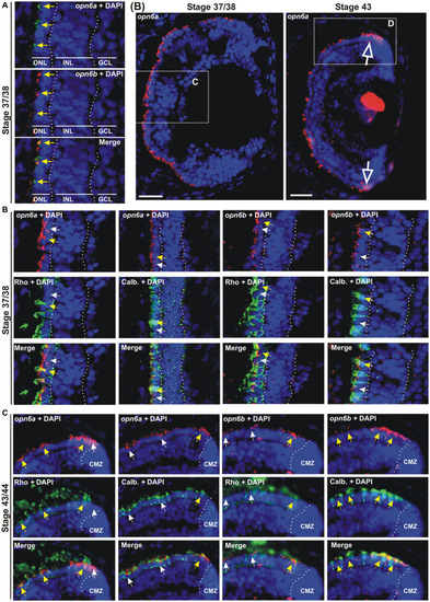
opn6a and opn6b colocalize in newly born photoreceptors at stage 37/38 but are differentially expressed in differentiated rods and cones at stage 43/44. (A–D) Fluorescent ISH with specific riboprobes against opn6a or opn6b on stage 37/38 (A,C) and 43/44 Xenopus laevis transverse retinal sections (B,D), followed by immunohistochemistry against a rod marker (rhodopsin; green) or a cone marker (calbindin; green) (C,D). DAPI (blue) stains nuclei. (A) Yellow arrows point to ONL cells that express both opn6a and opn6b. (B) Higher magnification of the dotted-line boxes are shown in bottom panels (C,D). The open white arrows point to opn6a+ cells that are adjacent to the ciliary marginal zone (CMZ). (C,D) White small arrows point to opn6a or opn6b expressing cells that are either not rhodopsin+ or not calbindin+, while yellow arrows indicate opn6a+ or opn6b+ cells that are also positive for rhodopsin or calbindin. GLC, ganglion cell layer; INL, inner nuclear layer; ONL, outer nuclear layer. Scale bar in (B): 100 μm.
|
