
FIGURE
Fig. 3
- ID
- ZDB-FIG-230912-24
- Publication
- Abu Bakar et al., 2023 - Embryonic mercury exposure in zebrafish: Alteration of metabolites and gene expression, related to visual and behavioral impairments
- Other Figures
- All Figure Page
- Back to All Figure Page
Fig. 3
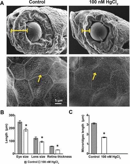
Fig. 3. Scanning electron microscopic (SEM) analyses of HgCl2–exposed zebrafish at 6 dpf. (A) Eye and lens size, retinal thickness (indicated by bar size), and microridges (arrow) in keratinocytes were examined. (B) Measurements of eye size, lens size, and retinal thickness. Embryonic exposure to HgCl2 caused a significant decreased in ocular structures. (C) Reduction in microridge length in keratinocytes in HgCl2–exposed zebrafish compared with control (mean ± SEM: *p < 0.05, n = 10 for each group). |
Expression Data
Expression Detail
Antibody Labeling
Phenotype Data
Phenotype Detail
Acknowledgments
This image is the copyrighted work of the attributed author or publisher, and
ZFIN has permission only to display this image to its users.
Additional permissions should be obtained from the applicable author or publisher of the image.
Full text @ Ecotoxicol. Environ. Saf.