FIGURE

Figure 3

ID
ZDB-FIG-230828-27
Publication
Staff et al., 2023 - Skin Extracellular Matrix Breakdown Following Paclitaxel Therapy in Patients with Chemotherapy-Induced Peripheral Neuropathy
Other Figures
All Figure Page
Back to All Figure Page
Figure 3

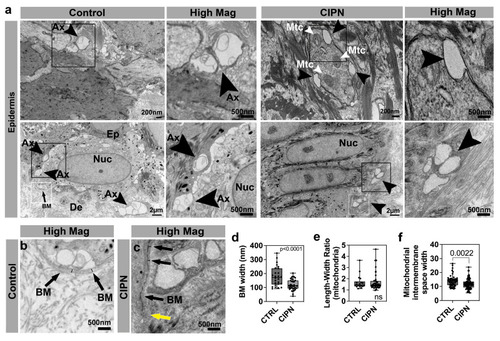
Epidermal changes in CIPN patients. (a) Intraepidermal nerve endings (arrowheads) are abundant in control subject skin whereas gaps are present in the CIPN patient skin. (b) High magnification of control skin shown in (a) depicting the basement membrane (arrows) and width demarcated by the black lines. (c) High magnification of CIPN patient skin shown in (a) depicting the basement membrane (black arrows) and width demarcated by the black lines. The yellow arrow points to the less dense region of the basement membrane. (d) Quantification of the basement membrane width (white boxes in a) shows a reduction in the CIPN patient skin. (e) Length–width ratio (LWR) of mitochondria in epidermal keratinocytes shows no differences between the control subjects and CIPN patients. (f) Mitochondrial intermembrane space reduction in CIPN patients. Abbreviations: Ax: Axon, BM: Basement membrane, De: Dermis, Ep: Epidermis, Mtc: Mitochondria, Nuc: Nucleus.

Expression Data

Expression Detail
Antibody Labeling
Phenotype Data

Phenotype Detail
Acknowledgments
This image is the copyrighted work of the attributed author or publisher, and ZFIN has permission only to display this image to its users. Additional permissions should be obtained from the applicable author or publisher of the image. Full text @ Cancers