FIGURE

Figure 2

ID
ZDB-FIG-230828-26
Publication
Staff et al., 2023 - Skin Extracellular Matrix Breakdown Following Paclitaxel Therapy in Patients with Chemotherapy-Induced Peripheral Neuropathy
Other Figures
All Figure Page
Back to All Figure Page
Figure 2

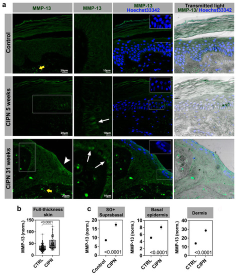
MMP-13 immunofluorescence staining in different skin compartments. Stratum spinosum and granulosum, SS + SG; stratum basale, SB; and dermis. (a) MMP-13 antibody staining in control (top panel) and two CIPN patients (middle panel at 5 weeks, and lower panel at 31 weeks post CIPN diagnosis). White boxes depict zoomed-in regions shown in the second column. MMP-13 positive cells are present in the basal (SB) layer (white arrows). The white arrowhead in the lower left panel points to MMP-13 positive cells in the SS + SG layer. The yellow arrows depict increased dermal expression in the CIPN skin. Insets with Hoechst33342 stain show darker nuclei in the CIPN patient skin compared with the control subject skin. The scale in unlabeled images is identical to the panel on the left. (b) Quantification of MMP-13 expression in full-thickness skin. (c) Quantification of MMP-13 expression in sub-compartments (SG + SS vs. SB vs. dermis).

Expression Data

Expression Detail
Antibody Labeling
Phenotype Data

Phenotype Detail
Acknowledgments
This image is the copyrighted work of the attributed author or publisher, and ZFIN has permission only to display this image to its users. Additional permissions should be obtained from the applicable author or publisher of the image. Full text @ Cancers