|
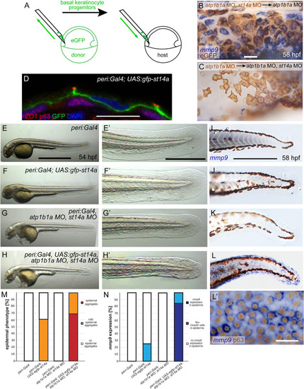
St14a functions in the periderm to activate signaling in the underlying basal layer. (A-C) Loss of st14a in basal cells is neither necessary nor sufficient to normalize mmp9 expression in basal cells of atp1b1a morphants. (A) Schematic of experimental set up in which ventral ectodermal cells either from atp1b1a, st14a double morphant or atp1b1a morphant donors were homotopically transplanted at 6 hpf into the same region of atp1b1a or atp1b1a, st14a double morphant hosts, respectively (atp1b1a MO, st14a MO>atp1b1a MO or atp1b1a MO>atp1b1a MO, st14a MO). Donor cells express Tg(Ola.Actb:Hsa.hras-egfp)-encoded membrane-tagged eGFP. Whole mount in situ hybridization for mmp9 (blue) and immunohistochemistry for eGFP (brown) of chimeric embryos at 58 hpf, showing that in atp1b1a MO, st14a MO>atp1b1a MO experiments, atp1b1a MO, st14a MO basal cells express mmp9 (B), and are thus not rescued although they lack functional Matriptase-1; whereas in atp1b1a MO>atp1b1a MO, st14aMO experiments, atp1b1a MO basal cells lack mmp9 expression (C), and are thus rescued although they contain Matriptase-1 (n = 6–8). (D-N) Re-introduction of st14a into peridermal cells of atp1b1a, st14a double morphants abrogates the rescue of basal cells, reflected by re-gained epidermal aggregate formation and enhanced mmp9 expression. (D) Immunofluorescence for eGFP-Matriptase1a (green), p63 as a nuclear marker for basal cells (red) and ZO1/Tjp1 as a marker for tight junctions in peridermal cells (red) on transverse section through the epidermis of a 48 hpf embryo transgenic for peri:Gal4zc1044a and UAS:gfp-st14afr58Tg, counterstained with DAPI (blue). Transgene-encoded Matriptase-1 is restricted to peridermal cells and localized at their basolateral membranes. (E-H’) Brightfield images of representative live 56 hpf embryos transgenic for peri:Gal4zc1044a or peri:Gal4zc1044a; UAS:eGFP-st14a controls (E,F) and injected with atp1b1a and st14a morpholinos (atp1b1a MO, st14a MO) (G,H); lateral views of entire embryos (E-H), and magnified views of tail region of same embryos (E’-H’). In contrast to atp1b1a MO, st14a MO embryos without transgenic re-introduction of st14a (G,G’), the atp1b1a MO, st14a MO embryos transgenic for peri:Gal4zc1044a; UAS:eGFP-st14a displays epidermal aggregates (H,H’), comparable to global atp1b1a single mutants (compare with Fig 1L’). (I-L’) Representative whole mount mmp9 in situ hybridizations, revealing re-gained strong mmp9 expression in basal cells of 72 hpf atp1b1a MO, st14a MO embryo transgenic for peri:Gal4zc1044a; UAS:eGFP-st14a (L,L’, the latter counterstained for the basal cell marker p63), but not in the atp1b1a MO, st14a MO embryos lacking transgene-driven peridermal st14a re-expression (K). (M) Quantification of epidermal phenotype of 56 hpf embryos with respective genotypes, as shown in (E-H) (n = 20–46). (N) Quantification of mmp9 in situ signal of 72 hpf embryos with respective genotypes, as shown in (I-L) (n = 12–38). Scale bars: 20 μm (B,L’), 10 μm (D), 500 μm (E), 100 μm (E‘,I).
|