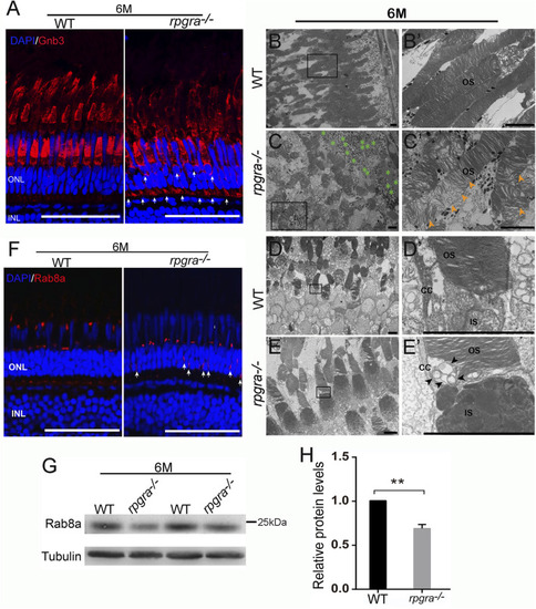
Abnormal ciliary trafficking in rpgra−/− zebrafish photoreceptors. (A) Retinal cryosections from WT and rpgra−/− zebrafish were labeled Gnb3 with specific antibodies at the ages of 6 mpf. Scale bars, 50 μm. (B) Well-maintained outer segments (OS) from WT retina. (B′) Enlarged image of the box in (B). Scale bars, 5 μm. (C) Disorganized and loosely arranged outer segment membrane disc in rpgra−/− zebrafish retina. Green asterisks, Lipid droplet. (C′) Enlarged image of the box in (C). The yellow arrows show a large number of loosely arranged outer segment membrane discs. Scale bars, 5 μm. (D) The complete structure of connecting cilia without the accumulation of abnormal objects in WT photoreceptor cells. (E) Obvious accumulation of transport vesicles in the connective cilia of the rpgra−/− photoreceptor cells. (D′, E′) Enlarged images of the boxes in (D, E). The black arrows indicate the accumulated vesicles. CC, connecting cilia; OS, outer segment; IS, inner segment; Scale bar, 5 μm. (F) Immunofluorescence showed that Rab8A protein in 6-month-old rpgra−/− zebrafish is mislocated in photoreceptor cells. The arrow marks the mislocated signals; Scale bar, 50 μm. (G) Protein levels of Rab8a were detected by Western blot at the age of 6mpf. (H) Statistical result of Rab8a protein expression level. The quantitative data of five independent experiments were statistically analyzed using a two-tailed Student’s t-test and shown as mean ± SD. **, p < 0.01.
|
