FIGURE 5
- ID
- ZDB-FIG-230617-28
- Publication
- Morsli et al., 2023 - A p21-GFP zebrafish model of senescence for rapid testing of senolytics in vivo
- Other Figures
- All Figure Page
- Back to All Figure Page
|
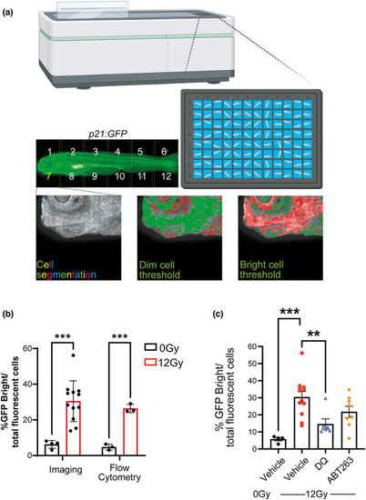
Senolytics reduces the number of p21:GFPBright cells. (a) Diagram representing automated imaging method for p21:GFP zebrafish using The Opera Phenix High‐Content Screening System. Tiled confocal photomicrographs from Opera Phenix microscope of 5 dpf p21:GFP zebrafish were acquired, and individual cells were segregated for analysis. The mean fluorescence intensity of individual cells was classified against a threshold set on the basis of level of fluorescence in wild‐type fish and non‐irradiated p21:GFP fish to identify the GFPBright population; (b) Percentage of GFPBright cells calculated by Opera Phenix High‐Content Imaging and Flow Cytometry analysis, as a proportion of total fluorescent cells in p21:GFP fish with or without irradiation. (c) Quantification of the proportion of GFPBright at 5 dpf in laterally oriented p21:GFP fish following irradiation at 2 dpf and treatment starting at 3 dpf with vehicle, dasatinib (D) plus quercetin (Q) or ABT263 (navitoclax). Data were examined with one‐way ANOVA with Tukey's multiple comparison's test. Data (from two independent experiments are presented as Mean ± SEM presented. *** |

