
Fig. 6
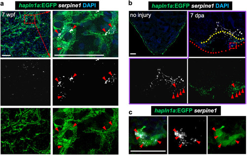
a Visualization of hapln1a:EGFP+ cells and in situ signals of serpine1 mRNA in whole-mount view of the ventricular surface of juvenile hapln1a:EGFP animals. Red dashed rectangle is enlarged. Red arrowheads indicate in situ signals, located in hapln1a+ cells. n = 6. Scale bar, 50 µm. b In situ of serpine1 mRNA in section views of adult injured hapln1a:EGFP hearts. Red arrowheads indicate in situ signals distributed in EGFP+ cells, which are near the area of coronary vessel regrowth. Yellow dashed lines represent surgery plane. Red dashed lines outline ventricular apex. Red rectangle area is enlarged in c. n = 5. Scale bar, 50 µm. c Representative image of serpine1 in situ signals located in EGFP+ cells in the wound area. Red arrowheads indicate in situ signals. Scale bar, 25 µm. |