
Fig. 1
- ID
- ZDB-FIG-230509-21
- Publication
- Nikolaou et al., 2022 - Cytoplasmic pool of U1 spliceosome protein SNRNP70 shapes the axonal transcriptome and regulates motor connectivity
- Other Figures
- All Figure Page
- Back to All Figure Page
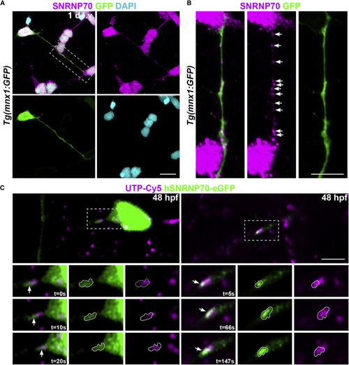
Figure 1. SNRNP70 localizes to axons (A) A representative image of a dissociated motor neuron at 1 day in vitro derived from Tg(mnx1:GFP) animals at 28 hpf. The sample was stained with anti-SNRNP70 to reveal the localization of the protein in neurites and DAPI to mark nuclei. Scale bars, 10 μm. (B) Inset showing a higher magnification of the motor axon in (A). Arrows indicate SNRNP70+ puncta within the GFP+ axon. Scale bars, 5 μm. (C) Time-lapse of neurons at 48 hpf sparsely labeled with hSNRNP70-eGFP. Top: images show GFP-labeled neurons co-expressing UTP-Cy5. Bottom: insets depict axon segments showing association between Cy5-RNA granules (magenta) and hSNRNP70-eGFP (green) signals (arrows indicate the same punctum during the imaging session with time shown in the bottom right corner of individual time points). In the single-channel images, the hSNRNP70-eGFP fluorescence signal has been outlined and overlayed in the UTP-Cy5 signal for a direct comparison. Representative examples from three independent experiments. Scale bars, 5 μm. See also Figure S1 and Video S1. |