
Figure 4.
- ID
- ZDB-FIG-230321-4
- Publication
- Canham et al., 2023 - EVA1A (Eva-1 Homolog A) Promotes Endothelial Apoptosis and Inflammatory Activation Under Disturbed Flow Via Regulation of Autophagy
- Other Figures
- All Figure Page
- Back to All Figure Page
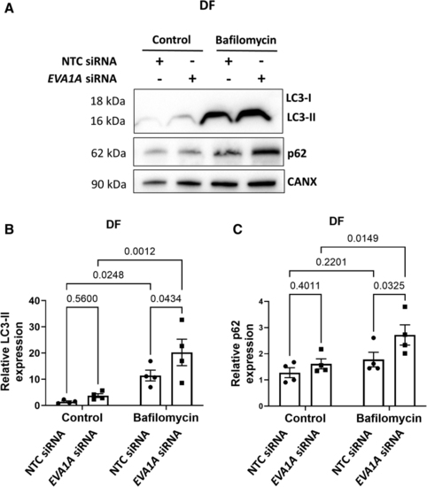
EVA1A reduces autophagic flux under disturbed flow. A through C, Human umbilical vein endothelial cells (HUVECs) were treated with EVA1A siRNA (small interfering RNA) or with nontargeting control (NTC) siRNA before exposing them to flow for 72 h using the orbital shaker system. To block autophagic flux, ECs were treated with 50 nM bafilomycin for the last 4 h of flow exposure. Control cells were treated with 0.05% dimethyl sulfoxide (DMSO). ECs were isolated from the disturbed flow (DF) region and expression levels of autophagy markers LC3-II (microtubule-associated protein 1 light chain 3-II) and p62 were assessed by Western blotting. Calnexin was used as a loading control. B and C, Graphs show protein levels of LC3-II and p62 normalized to CANX (calnexin; n=4). Data are presented as means±SE of the mean. Differences between groups were analyzed using a 2-way ANOVA (B and C) with Tukey post hoc test and P values are shown in the graphs. |