
Figure 3.
- ID
- ZDB-FIG-230321-3
- Publication
- Canham et al., 2023 - EVA1A (Eva-1 Homolog A) Promotes Endothelial Apoptosis and Inflammatory Activation Under Disturbed Flow Via Regulation of Autophagy
- Other Figures
- All Figure Page
- Back to All Figure Page
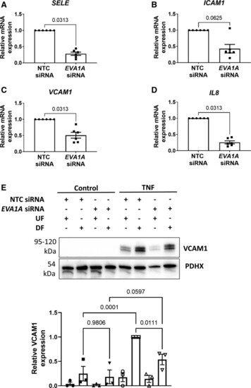
EVA1A (eva-1 homolog A) promotes endothelial permeability and inflammatory activation under disturbed flow. A through E, Human umbilical vein endothelial cells (HUVECs) were treated with EVA1A siRNA (small interfering RNA) or with nontargeting control (NTC) siRNA before exposing to flow for 72 h using the orbital shaker system. A through D, ECs were isolated from the disturbed flow (DF) region, and mRNA expression of E-selectin (SELE), intercellular adhesion molecule 1 (ICAM1), vascular cell adhesion molecule (VCAM1), and interleukin 8 (IL8) were measured by quantitative real-time polymerase chain reaction (qRT-PCR; n=6), using HPRT as a housekeeping gene. E, ECs were stimulated with TNF (tumor necrosis factor; 10 ng/mL) for the last 4 h of flow. VCAM1 protein expression was assessed in TNF-stimulated and nonstimulated (control) ECs isolated from the DF and undisturbed flow (UF) regions by Western blotting. Graph shows VCAM1 protein levels normalized to PDHX (pyruvate dehydrogenase complex component X; n=3). A through E, Data are presented as means±SEM and normalized to control. Differences between groups were analyzed using a nonparametric Wilcoxon test (A–D) or a 2-way ANOVA with Tukey post hoc test (E). P values are shown in the graphs. |