
Fig 4
- ID
- ZDB-FIG-230315-54
- Publication
- Xie et al., 2023 - Ependymal polarity defects coupled with disorganized ciliary beating drive abnormal cerebrospinal fluid flow and spine curvature in zebrafish
- Other Figures
- All Figure Page
- Back to All Figure Page
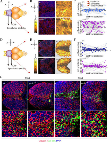
Loss of Ccdc57 results in ependymal cell polarity defects.
(A, B) Distribution pattern of ependymal cells in 3-months-old wild type and ccdc57 mutant as indicated with tight junction marker Claudin-5 staining. Nuclei were counterstained with DAPI in blue. The displayed region of ependymal epithelia was indicated in panel A. Heatmap showing the relative size of each ependymal cells as displayed in the confocal images. (C) Statistical analysis showing orientation of the midline ependymal cells along the anterior–posterior (A–P) axis in wild type and ccdc57 mutant. The angles between the longer axis of each midline ependymal cells and the A–P axis were used to evaluate cell polarity with 0 degree indicating horizontal cell. (D, E) Distribution pattern of ependymal cells in the brains of 17 dpf wild type and ccdc57 mutant. (F) Statistical analysis showing orientation of the midline ependymal cells in wild type and mutant as indicated. (G) Confocal images showing the ependymal layer labeled with Claudin-5 (red) and glycylated tubulin (green) antibodies in 25 and 30 dpf wt and ccdc57 mutants. Enlarged views of the boxed regions are displayed in the bottom. Nuclei were counterstained with DAPI. Scale bars: 25 μm in panel B; 50 μm in panel E; 50 μm in panel G. The data underlying the graphs shown in the figure can be found in S1 Data. |
| Antibodies: | |
|---|---|
| Fish: | |
| Anatomical Terms: | |
| Stage Range: | Days 14-20 to Adult |
| Fish: | |
|---|---|
| Observed In: | |
| Stage Range: | Days 14-20 to Adult |