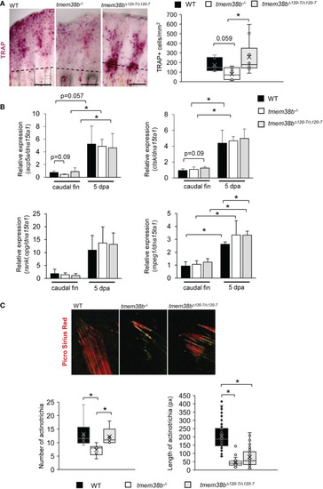
Osteoclast analysis during caudal fin regeneration in tmem38b mutants. (A) Representative images of TRAP staining of caudal fins of WT and tmem38b mutants at 5 dpa (WT n = 7, tmem38b-/- n = 9, tmem38bΔ120-7/Δ120-7 n = 10). In WT, osteoclasts were present in the regenerate. Almost no TRAP activity could be detected in tmem38b-/- , while in tmem38bΔ120-7/Δ120-7 it seemed to be mostly localized along the regenerating fin rays. TRAP+ cells were significantly reduced in tmem38b-/- respect to the other two groups. (B) Relative expression of bone resorption-related markers acp5a and ctsk, of the macrophage marker mpeg1 and rankl/opg ratio in amputated and 5 dpa caudal fin (WT n = 3, tmem38b-/- n = 3, tmem38bΔ120-7/Δ120-7 n = 3). All markers were increased after amputation. mpeg1 was significantly overexpressed in tmem38bΔ120-7/Δ120-7 compared to WT at 5 dpa.*: p < 0.05. (C) Representative images of picro sirius red staining of actinotrichia in caudal fins of WT and tmem38b mutants (WT n = 3, tmem38b-/- n = 3, tmem38bΔ120-7/Δ120-7 n = 3). tmem38b-/- revealed a reduced number of actinotrichia respect to WT and tmem38bΔ120-7/Δ120-7 , while the length of actinotrichia was reduced in both mutants compared to WT. *p < 0.05.