
FIGURE 2
- ID
- ZDB-FIG-230110-131
- Publication
- Scott et al., 2022 - Aerobic glycolysis is important for zebrafish larval wound closure and tail regeneration
- Other Figures
- All Figure Page
- Back to All Figure Page
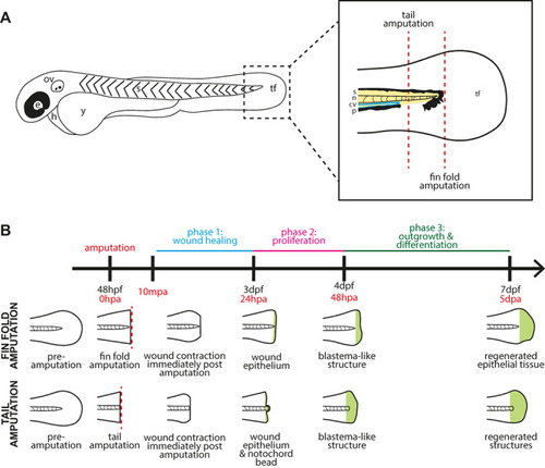
Schematic diagrams of the fin and tail amputation models of zebrafish embryo regeneration. (A) Schematic drawing of a two‐day post fertilisation zebrafish embryo. e: eye, ov: otic vesicle, h: heart, y: yolk, s: somites, tf: tail fin. Dashed box indicates the area shown enlarged in the box to the right, which depicts the tail region with amputation planes for fin fold and tail amputations (dashed red lines). A fin fold amputation site is positioned just distal to the tip of the notochord and excision is limited epithelial tissue and mesenchymal cells, while a tail amputation is oriented using the pigment gap and circulatory loop of the caudal vein, severing notochord, neural tube, and muscle in addition to epithelial tissue. s: somites (yellow), n: notochord (pink), cv: caudal vein (blue), p: pigment (black), tf: tail fin fold. (B) General timeline and schematic illustration of phases of regeneration following amputation. Embryos are imaged at various time points depending on the specific experiment between ten minutes post amputation (10mpa) and full regeneration at five days post amputation (5dpa). |