
Fig. 5
- ID
- ZDB-FIG-221221-6
- Publication
- Glasauer et al., 2021 - DNA template strand segregation in developing zebrafish
- Other Figures
- All Figure Page
- Back to All Figure Page
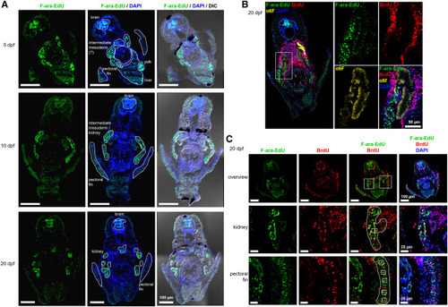
Figure 5. Label-retaining cells in the zebrafish mid-body region (A) Zygotes were microinjected with 5.7 pmol F-ara-EdU, and fish were grown to 5, 10, or 20 dpf. Fixed and sectioned zebrafish were stained with Alexa Fluor 488 azide and total DNA was counterstained using DAPI. (B) Same treatment as in (A), but sections were additionally stained with the kidney marker alpha6F. (C) Same treatment as in (A), but fish were additionally incubated in 10 mM BrdU for 24 h prior to fixation. Sister cell pairs are marked by white boxes in the increased magnification images (middle and bottom row). Transverse sections through the mid-body region are shown. |