
FIGURE
Fig. 4
- ID
- ZDB-FIG-221028-19
- Publication
- Cacialli et al., 2022 - Synergistic prostaglandin E synthesis by myeloid and endothelial cells promotes fetal hematopoietic stem cell expansion in vertebrates
- Other Figures
- All Figure Page
- Back to All Figure Page
Fig. 4
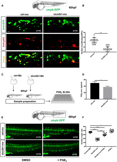
The defect in HSPCs proliferation in slco2b1‐deficient embryos can be rescued by PGE2 treatment
Source data are available online for this figure. |
Expression Data
| Gene: | |
|---|---|
| Fish: | |
| Condition: | |
| Knockdown Reagent: | |
| Anatomical Term: | |
| Stage: | Pec-fin |
Expression Detail
Antibody Labeling
Phenotype Data
| Fish: | |
|---|---|
| Condition: | |
| Knockdown Reagent: | |
| Observed In: | |
| Stage: | Pec-fin |
Phenotype Detail
Acknowledgments
This image is the copyrighted work of the attributed author or publisher, and
ZFIN has permission only to display this image to its users.
Additional permissions should be obtained from the applicable author or publisher of the image.
Full text @ EMBO J.