
Fig. 7
- ID
- ZDB-FIG-221011-12
- Publication
- Vandestadt et al., 2021 - RNA-induced inflammation and migration of precursor neurons initiates neuronal circuit regeneration in zebrafish
- Other Figures
- All Figure Page
- Back to All Figure Page
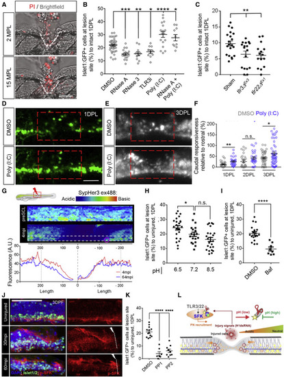
Figure 7. An RNA-induced inflammatory response mediates tissue regeneration and precursor neuron recruitment (A) Maximum projection of the injury site at 2 and 15 min post lesion (MPL), showing propidium iodide (PI) uptake in cells at the injury site. Lesion shown with white line. (B) Quantification of isl1:GFP+ cells at 1 DPL relative to control (DMSO) following RNase A, RNase III, TLR3i, poly(I:C), or RNase A, followed by poly(I:C) treatment (C) Quantification of isl1:GFP+ cells at 1 DPL relative to control siblings in tlr3 and tlr22 mutants with deleted RNA-binding domains. (D) Maximum projection of poly(I:C)-treated fish at 1 DPL showing a significant boost in the recruitment of isl1:GFP+ cells to the lesion site. (E and F) Quantification of neural activity using GCaMP6sΔF following poly(I:C) treatment showing enhanced recruitment of neurons and improved neural activity downstream of the injury compared with control. (G) Measurement of local pH changes in response to injury using the in vivo pH indicator Sypher3. The white outline shows region of analysis in the bottom panel and the red circle shows cells analyzed in Figures S7J and S7K. Bottom panel; intensity plot for ventral cell population at 4 (red) and 64 (blue) min postinjury (MPI) showing a rapid drop in pH in proximity to injury. (H) Quantification of isl1:GFP+ cells following treatment with different pH in media showing that an acidic environment after injury increases the number of isl1:GFP+ cells in the lesion site compared with neutral pH. (I) Quantification of isl1:GFP+ cells at injury site 1 DPL, following bafilomyocin-A1 (Baf) treatment, showing a reduction in isl1:GFP+ cell recruitment in Baf-treated fish compared with vehicle control. (J) Representative maximum projection images of pSFK-immunostained isl1:GFP in intact and injured larvae. (K) Quantification of isl1:GFP+ cells at 1 DPL following the inhibition of SFK signaling using PP1 or PP2. (L) Scheme showing that the acidic cellular environment created by injury facilitates interaction of dsRNA with TLRs, which, in turn, activates SFK phosphorylation and promotes rapid migration of isl1:GFP+ cells to the lesion. Error = SEM, n.s., not significant, ∗p < 0.05, ∗∗p < 0.01, ∗∗∗p < 0.001, ∗∗∗∗p < 0.0001 Scale bar, 50 μm, dorsal up and rostral left. |
Reprinted from Developmental Cell, 56, Vandestadt, C., Vanwalleghem, G.C., Khabooshan, M.A., Douek, A.M., Castillo, H.A., Li, M., Schulze, K., Don, E., Stamatis, S.A., Ratnadiwakara, M., Änkö, M.L., Scott, E.K., Kaslin, J., RNA-induced inflammation and migration of precursor neurons initiates neuronal circuit regeneration in zebrafish, 2364-2380.e8, Copyright (2021) with permission from Elsevier. Full text @ Dev. Cell