
Fig. 3
- ID
- ZDB-FIG-220926-3
- Publication
- Jussila et al., 2022 - Live imaging and conditional disruption of native PCP activity using endogenously tagged zebrafish sfGFP-Vangl2
- Other Figures
- All Figure Page
- Back to All Figure Page
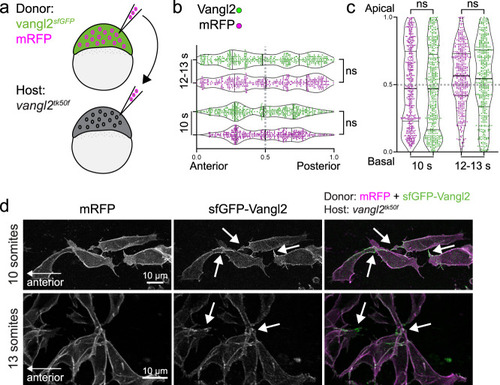
Planar polarized enrichment of Vangl2 is controlled by cell non-autonomous PCP signalling.
a A schematic illustrating transplantation of membrane-RFP-labelled vangl2sfGFP donor cells into vangl2tk50f loss-of-function host embryos, at sphere stage. b, c Distribution of the brightest sfGFP-Vangl2 and membrane-RFP spots along anterior-posterior (g) and apical-basal (h) axes of vangl2sfGFP neuroepithelial cells transplanted into a vangl2tk50f mutant host, quantified at two consecutive stages of neural tube morphogenesis. Data from multiple transplanted vangl2sfGFP cells was pooled (10 somites, n = 7; 12-13 somites, n = 9). Statistical analysis was performed using a two-tailed Mann-Whitney test. Thicker line shows median, and thinner lines first and third quartile. Source data are provided as a Source Data file. d Representative confocal images of membrane-RFP and sfGFP-Vangl2 localization in vangl2sfGFP neuroepithelial cells within vangl2tk50f mutant hosts, at 10-somite and 13-somite stages of development, as quantified in b, c. Maximum intensity projections are shown. Arrows point at Vangl2 enrichment on cell protrusions. Anterior is to the left in all images. |