
FIGURE
Fig. 5
- ID
- ZDB-FIG-220905-21
- Publication
- Waldmann et al., 2021 - The role of Gdf5 in the development of the zebrafish fin endoskeleton
- Other Figures
- All Figure Page
- Back to All Figure Page
Fig. 5
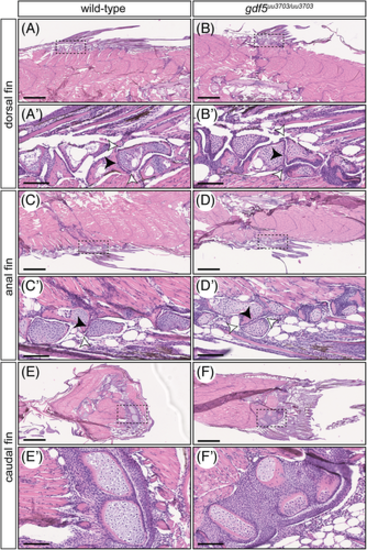
Median fin histology of adult wild-type and gdf5uu3703/uu3703 zebrafish reveals intact joints between distal and proximal radials and normal maturation of the skeletal elements. Sagittal sections from 60 dpf of wild-type (A,A,C,C′,E,E′) and gdf5uu3703/uu3703 (B,B′,D,D′,F,F′) zebrafish median fins stained with Hematoxylin and Eosin (HE). Anterior to the left, dorsal to the top. Dashed boxes in A-F mark magnified region in A′-F′. (A,A′,B,B′) Dorsal fin; (C,C′,D,D′) anal fin; (E,E′,F,F′) caudal fin. (A′-D′) White arrowheads indicate joint capsule, black arrowheads indicate articular cartilage. All joints are intact and do not display any differences when compared to wild-type. (E-F,E′-F′) Caudal fin skeletal elements display no apparent delay in chondrocyte maturation when compared to wild-type. Scale bars: 500 μm (A-F), 100 μm (A′-F′)
|
Expression Data
Expression Detail
Antibody Labeling
Phenotype Data
| Fish: | |
|---|---|
| Observed In: | |
| Stage: | Days 45-89 |
Phenotype Detail
Acknowledgments
This image is the copyrighted work of the attributed author or publisher, and
ZFIN has permission only to display this image to its users.
Additional permissions should be obtained from the applicable author or publisher of the image.
Full text @ Dev. Dyn.