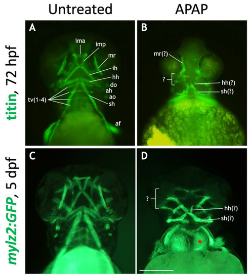
Many craniofacial muscles are absent or disorganized in APAP-treated larvae. (A,B): Ventral views of 72-hpf embryos processed for titin immunohistochemistry to label muscles. In an untreated embryo (A), ventral muscle types are in focus and labeled according to type. In an APAP-treated embryo (B), developing muscles are present but disorganized. (C,D): Ventral views of live-imaged, 5-dpf mylz2:GFP larvae which express GFP in skeletal muscles. Compared to an untreated larva (C), an APAP-treated larva (D) has a reduced number of misarranged craniofacial muscles. Due to missing musculoskeletal elements from branchial arches three though seven, the underlying anterior somites are visible (red asterisk in (D)). The defects in both titin-stained and mylz2:GFP larvae are variable, but the most common arrangements are shown here. Abbreviations (after Schilling and Kimmel 1997): aboral fin, af; adductor hyoideus, ah; adductor opercula, ao; dilator opercula, do; hyohyoideus, hh; interhyoideus, ih; intermandibularis anterior, ima; intermandibularis posterior, imp; medial rectus, mr; sternohyoideus, sh; transversus ventralis, tv. Scale bar (in (D)) for (A–D), 200 µm.
|
