FIGURE

Figure 4

ID
ZDB-FIG-220801-56
Publication
Lin et al., 2022 - Quantification of Idua Enzymatic Activity Combined with Observation of Phenotypic Change in Zebrafish Embryos Provide a Preliminary Assessment of Mutated idua Correlated with Mucopolysaccharidosis Type I
Other Figures
All Figure Page
Back to All Figure Page
Figure 4

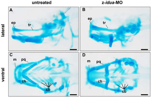
Knockdown of z-idua caused defective head cartilage in zebrafish embryos. Using alcian blue staining to detect the head cartilage of zebrafish embryos at 120 hpf. Untreated embryos (control) and embryos injected with antisense morpholino oligonucleotides (MO) complementary with z-idua (z-idua-MO) were studied. (A,B) Lateral view; (C,D) Ventral view; (A,C) Untreated control embryos; and (B,D) z-idua-MO injected embryos. Loss of function of z-Idua caused deformed cranial cartilage and decreased number of cartilage cells. cb, ceratobranchial; ch, ceratohyal; ep, ethmoid plate; m, Meckel’s cartilage; pq, palatoquadrate; tr, trabecular plate. Scale bar: 100 μm.

Expression Data

Expression Detail
Antibody Labeling
Phenotype Data

Phenotype Detail
Acknowledgments
This image is the copyrighted work of the attributed author or publisher, and ZFIN has permission only to display this image to its users. Additional permissions should be obtained from the applicable author or publisher of the image. Full text @ J Pers Med